Dr. Geoffrey L. Sas focuses on treatment planning decisions and the best evidence to properly restore endodontically treated teeth
 The primary objective of endodontic therapy is to prevent and treat apical periodontitis.1 To effectively achieve this goal, proper cleaning and shaping of the canals, irrigation, and a coronal seal are essential. The objectives of restorative dentistry are to properly restore teeth to function, comfort, and in specific cases, esthetics. Although the materials and methods of both treatment modalities have changed, the ultimate goals have remained constant. The relationship between endo-dontic treatment and restorative dentistry has been established. However, the concepts and related treatment plans have been contentious. With the increasing publicity regarding “implant” dentistry, there is an emphasis on evaluating the restorability of teeth prior to endodontic treatment. It is not beneficial to the patient if the root canal therapy (RCT) is successful, but the tooth ultimately fails. With the advancement of implant dentistry, diseased teeth that previously may have had root canal therapy and a crown now may be replaced with implants, provided the long-term restorability is in question or is dictated by the overall treatment plan. This article focuses on treatment planning decisions and the best evidence to properly restore endodontically treated teeth.
Long-term success of endodontically treated teeth is dependent on the ensuing restorative treatment.2 Microorganisms that may cause apical periodontitis and contamination of the root canal system during or after endodontic therapy can alter the ultimate success of the diseased tooth.3 The growth of bacteria through the exposure of gutta percha to saliva results in endotoxin at the apex within days of endodontic treatment. Delays in final restoration after completion of RCT have been indicative of lower success rates.4
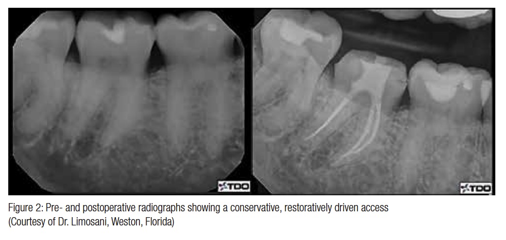
 There are differing opinions regarding endodontic access and its role in restorative dentistry. Many “Coke™ bottle” preparations used in the past unnecessarily removed cervical dentin2 (Figure 1). Access designs should focus on conserving as much tooth structure as possible without compromising the RCT (Figure 2). Adhesive materials used in the coronal restoration provide an immediate seal and strengthening of the tooth. A major benefit of adhesive dentistry is that it does not solely rely on mechanical retention, and therefore, tooth structure can be preserved.5 Notwithstanding the numerous advantages to bonding within the root canal system, there are also limitations such as the geometry of the canal. The ratio of bonded to unbonded surfaces is called the configuration factor or “C” factor. A higher percentage of unbonded surfaces results in less stress on the bonded surfaces from polymerization contraction. A Class IV preparation has a C factor of less than 1:1, which is favorable compared to the root canal system that may be as high as 100:1.6 With an unfavorable geometry, it is not possible to achieve an ideal gap-free interface between the gutta percha and adhesive materials, and therefore, the long-term seal could be altered. As well, it is technically challenging to apply primer and adhesive deep in the root canal system.
 The principle of cuspal coverage is a consistent factor throughout the literature and is the most consistent factor when predicting survivability of root canal (RC) treated teeth. Aquilino and Caplan showed that when tooth type and presence of caries at the time of access were controlled, at a 9-year follow-up exam, teeth with cuspal coverage had a 6 times greater survival rate than teeth without cuspal coverage.7 Aquilino and Caplan concluded that although treatment recommendations should be made on an individual basis, the associations between crowns and the survival of RC treated teeth should be recognized.7 Coronal tooth structure should be preserved just as much as radicular. For teeth that require posts as part of their coronal restoration, no additional dentin should be removed beyond what is necessary for root canal treatment. As an example, if the canal is prepared to a 0.04 preparation, a 0.04 tapered post should “fall” right in without mechanically preparing the canal to fit the post. There is near consensus that the ferrule effect is very important when treatment planning a single diseased tooth. Ferrule is cervical tooth structure that provides retention and resistance form to the restoration, which prevents fracture. Ferrule is best when it is at least 1.5 mm-2 mm or more and is important to long-term success when a post is used.8 If the height of the remaining tooth structure does not have adequate ferrule, options may include crown lengthening, orthodontic extrusion, or extraction and replacement.
The function of a post is strictly to retain a core in a tooth with extensive loss of tooth structure.9 Although custom cast posts or prefabricated metal posts have become the standard for decades, in recent years, fiber-reinforced composite posts are increasingly more prevalent. Placing posts comes with inherent risks such as disturbing the root canal filling material, which may lead to microleakage, increased risk of perforation, and iatrogenically removing tooth structure. The RC system should never be shaped to fit posts, and no instrument should be used in a canal unless it is intended to shape the canal for its endodontic obturation. Although metal posts do not reinforce the strength of the root structure, there is increasing evidence that fiber posts may increase resistance to fracture.10 The concept of a fiber post is that it has a modulus of elasticity similar to that of dentin and therefore can absorb more impact force and distribute force better than more rigid metal posts. As well, if failure occurs in a fiber post, the results are less severe.11 There are also esthetic advantages to using nonmetallic posts, particularly for anterior abutments.
Retention of posts is directly proportional to the length of the post. Several concepts have been suggested for passive fitting posts, such as ensuring that the post is at least apical to the crest of the alveolar bone, or at least equal to the crown height. When placing a post, it is important to maintain the endodontic seal. To maintain a long-term seal, 4 mm-5 mm of gutta percha (GP) is superior compared to 2 mm-3 mm.12 Hand instruments, rotary instruments, and heat can be used to remove GP without disrupting the apical seal. Goldfein, et al., confirmed that when a rubber dam was used during post placement, there was a significantly lower chance of developing a periapical lesion at the 2.7-year mark.13
A primary objective of endodontic therapy is to establish an adequate seal with the root canal filling material, as coronal microleakage is a leading cause of endodontic failure. The current trend of “temporizing” with cotton and cavit or another temporary material following endodontic treatment can result in various complications. First, patients may not return to their restorative dentist in a timely manner, and thus the provisional coronal restoration will rapidly break down and potentially cause microleakage. Placing an immediate core at the time of endodontic obturation is recommended to further the coronal seal, which is an integral part of endodontic therapy. The clinician’s knowledge of the canal angulations, anatomy, and curvature is greatest at the time of obturation, which makes that point the optimal time to place the buildup. Because the rubber dam is already present, the immediate buildup becomes an extension rather than an invasion of the endodontic seal. Ray and Trope evaluated the relationship between the quality of the coronal restoration and the quality of the root canal filling by examining the radiographs of endodontically treated teeth.14 They observed that a combination of good restorations and good endodontic treatments resulted in the absence of periapical inflammation in 91.4% of the teeth examined, whereas poor restorations and poor endodontic treatments resulted in the absence of periradicular inflammation in only 18.1% of teeth. Furthermore, where poor endodontic treatments were followed by good permanent restorations that appeared radiographically sealed, the resultant success rate was 67.6%. Consequently, Ray and Trope concluded that apical periodontal health depended significantly more on the coronal restoration than on the technical quality of the endodontic treatment.14 Mavec, et al., evaluated the bacterial microleakage of the remaining gutta percha in teeth prepared for a post space with and without the use of an intracanal glass ionomer cement barrier. They discovered that the length of time between obturation and placement of the permanent restoration is critical to prevent recontamination of the remaining apical gutta percha.15 In this study, Vitrebond™ (3M) proved an acceptable intracanal barrier material and should provide a superior secondary seal for the temporary coronal restoration. In conclusion, the application of a combined endodontic seal/buildup procedure in a timely manner combined with an adequate ferrule effect will significantly improve the long-term success of endodontic and restorative care.
This article was originally published in Oral Health Journal (reprinted with permission).
References
- Siqueira JF. Aetiology of root canal treatment failure: why well-treated teeth can fail. Int Endod J. 2001;34(1):1-10.
- Ree M, Schwartz RS. The endo-restorative interface: current concepts. Dent Clin N Am. 2010;54(2):345-374.
- Kakehashi S, Stanley HR, Fitzgerald RJ. The effects of surgical exposure of dental pulps in germ-free and conventional laboratory rats. Oral Surg Oral Med Oral Pathol. 1965;20:340-349.
- Alves J, Walton R, Drake D. Coronal leakage: endotoxin penetration from mixed bacterial communities through obturated, post-prepared root canals. J Endod. 1998;24(9):587-591.
- Van Meerbeek B, De Munck J, Yoshida Y, Inoue S, Vargas M, Vijay P, Van Landuyt K, Lambrechts P, Vanherle G. Buonocore memorial lecture. Adhesion to enamel and dentin: current status and future challenges. Oper Dent. 2003;28(3):215-235.
- Carvalho RM, Pereira JC, Yoshiyama M, Pashley DH. A review of polymerization contraction: the influence of stress development versus stress relief. Oper Dent. 1996;21(1):17-24.
- Aquilino SA, Caplan DJ. Relationship between crown placement and the survival of endodontically treated teeth. J Prosthet Dent. 2002;87(3):256-263.
- Stankiewicz N, Wilson P. The ferrule effect. Dent Update. 2008;35(4):227-228.
- Goodacre CJ, Spolnik KJ. The prosthodontic management of endodontically treated teeth: a literature review. Part 1. Success and failure data, treatment concepts. J Prosthodont. 1994;3(4):243-250.
- Naumann M, Preuss A, Frankenberger R. Reinforcement of effect of adhesively luted fiber reinforced composite versus titanium posts. Dent Mater. 2007;203(2):138-144.
- Butz F, Lennon AM, Heydecke G, Strub JR. Survival rate and fracture strength of endodontically treated maxillary incisors with moderate defects restored with different post-and-core systems: an in vitro study. Int J Prosthodont. 2001;14(1):58-64.
- Madison S, Wilcox, LR. An evaluation of coronal microleakage in endodontically treated teeth. Part III. In vivo study. J Endod. 1998;14(9):455-458.
- Goldfein J, Speirs C, Finkelman M, Amato R. Rubber dam use during post placement influences the success of root canal-treated teeth. J Endod. 2013;39(12):1481-1484.
- Ray HA, Trope M. Periapical status of endodontically treated teeth in relation to the technical quality of the root canal filling and the coronal restoration. Int Endod J. 1995;28(1):12-18.
- Mavec J, McClanahan SB, Minah GE, Johnson JD, Blundell RE Jr. Effects of an intracanal glass ionomer barrier on coronal microleakage in teeth with post space. J Endod. 2006;32(2); 120-122.
Stay Relevant With Endodontic Practice US
Join our email list for CE courses and webinars, articles and more..


