Dr. L. Stephen Buchanan offers his view of an innovative imaging technology
 A seldom addressed issue is the postural challenge of using operating microscopes in practice. This is probably because the advent of dental microscopes so dramatically improved dentists’ posture from the days of loupe magnification because they required dentists to sit more upright and to angle their patients’ heads instead of their own as they visualized the operative field. Better that patients have a neck ache for a couple of days after visiting the dentist, than the dentist becoming disabled by doing the same thing every day for years.
Despite this improvement, the ergonomic stress delivered to dentists using microscopes is still quite significant, as this limited range of position requires them to sit in static positions for hours, every day of practice. Microscopes used in dental practice have an inherently limited range of positioning due to the requirement that wherever the microscope is positioned over the patient’s face, the dentist must be able to look into the binocular eyepieces — a requirement that limits the microscope’s range of angulation to about 15°.
The muscle fatigue attendant to static positioning during practice is no different than that delivered to our patients who constantly hold or press their teeth together all day and all night long — the result is myofascial pain emanating from dentists’ backs and necks. I know this from personal experience, having had three spinal procedures to alleviate pinched sciatic nerves. After a day of intense practice with a microscope, I felt like my back and neck belonged to a 1,000-year-old endodontist.
The MoraVision™ 3D camera system — invented by Dr. Assad Mora, a prosthodontist in my home town of Santa Barbara — has changed all of that because with his imaging device, my view line is no longer posture-dependent. Now that I use his 3D camera system instead of a microscope, I can move at any and all times during procedures, and my back and neck feel as fresh at 6:30 p.m. as they did at 8:30 a.m.
Clinicians can now sit in any position, or better yet, in many positions during procedures because they only need to have a sight line to the 3D monitor on the wall of their operatories, eliminating the stress of static positioning. Not only does this imaging system help the operator, it also allows assistants to see the field exactly the same, an accomplishment that previously required them to also hold static positions at assistant’s scopes attached to the dentist’s scope. No longer will the image of a dentist and assistant huddled up to the binoculars of a microscope be the sine qua non of top-flight dental practices.
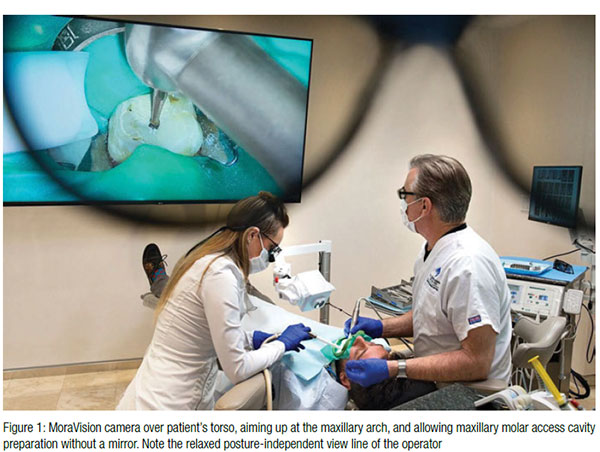
MoraVision™ takes vision ergonomics from a fixed, extremely limited line of sight that is totally dependent on the postural limitations of the operator, to a highly variable and posture-independent line of sight. Because this magnification device has been divorced from the operator’s face and the binocular eyepieces, the range of position is now a 180° hemisphere over the patient’s face, remarkably allowing access cavities and tooth preparations to be cut without a mirror when the camera is placed over the patient’s torso aimed up at their maxillary arch.
During the use of hand-held or rotary files on mandibular teeth, rather than dealing with the severe parallax errors that typically occur when sitting in a 12 o’clock position, the MV camera can be placed to either side of the mandibular tooth so the sight line is exactly orthogonal to the file, and the clinician sees the stop dead-on as it meets the reference point. Surprisingly, with this device, my hand-eye coordination remains intact even when the camera is aimed from these unusual angles.
 There have been other camera systems that displayed a dental operating field image to a monitor, such as Karl Storz’s EXOscope, and while they have delivered a greater depth of field than microscopes, the resulting 2D image typically requires months of practice to accommodate — similar to the time needed for endoscopic medical surgeons to train up to working without the third Z-plane dimension in their minimally-invasive procedures. The third dimension delivered by MoraVision’s stereoscopic twin cameras means that the learning curve in using this device is short and very intuitive — much less than I needed to become adept with microscopes. For most clinicians, the first time they view a patient’s mouth with this 3D system and enter the operative field with hands and instruments, it is a very obvious and natural thing to place them exactly where they need to be. The three-dimensionality of this device is the game changer; it makes all the difference.
 Why now and not before? Basically, like all disruptive technology, many different advancements must arrive and work together for its potential to be unleashed. Until 3D monitors were made to display the split-second representation needed by gamers, also a requirement for 3D dentistry, the small lag time between hand movement and depiction on the screen required some time to master and made it awkward to use. On the value proposition side of the equation, the price of 1080p camera systems finally came to a price point that delivers a good value for clinicians, when comparing the $38,500 cost of MoraVision to the $70,000 price of a ProErgo Zeiss microscope.
I have used this system for nearly a year during my live demonstrations for courses at my training center, Dental Education Laboratories, and the response has been amazement and fascination. Instead of a mirror view of the upper first molar I am treating, they get a straight-on, full view of the procedure. Beyond the obvious advantages of 3D presentation for live demos — one of which I did for last year’s ADA Session in Washington, DC — is the recording capabilities of the device. It ports directly into my TDO Chart where I can save still images or video in 2D and/or 3D format.
It’s a mind bender, but I am predicting that 3D will take implant surgery and general dentistry in a way microscopes never did because of these significant advantages:
- Depth of field
- Width of field
- Foot-controlled zoom and focus
- Its 180° hemisphere of positioning
- Its posture-independence and the improved ergonomics that it delivers
- Its ridiculously small size compared to microscopes
- Its ease of installation (Lift the edge of your chair; slide the pole support underneath; you are installed)
- Cost
- Simplicity and ease of clinical documentation
- Last but not least — its freakish cool factor
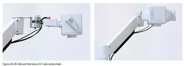
Dentists who have gone through the challenge of learning to use microscopes may be skeptical about this new inflection point in dental visualization. Dentists, specifically those who found microscopes to be unwieldy in general dentistry and implant surgery, finally have the light and magnification they need, elegantly presented in a manner that makes its use simple and intuitive when working in a wider field than an access cavity or retrograde surgical field. What would it be like to have perfect light and magnification with a depth of field that extends from third molars to incisors, installable in 20 minutes — all in a 5” cube? I’ve been using them since I sold my ProErgo scopes and, as usual, the view up front is the best.
Visit dentalcadre.com for information.
Stay Relevant With Endodontic Practice US
Join our email list for CE courses and webinars, articles and more..
