Editor’s intro: This article explores how various rotary systems work in ovoid canals. The complete removal of the root obturation material is vital to the effective disinfection of the root canal.
Drs. Aline Yumi Mukai, Ana Grasiela da Silva Limoeiro, Alexandre Sigrist De Martin, Augusto Shoji Kato, Carlos Eduardo da Silveira Bueno, Daniel Guimarães Pedro Rocha, Rina Andrea Pelegrine, and Carlos Eduardo Fontana discuss different systems for the removal of the obturation material
Abstract
Introduction
During nonsurgical endodontic retreatment, the complete removal of the previous filling material is necessary to facilitate proper cleaning, disinfection, and reintervention of the root canal system. This study aimed to evaluate the effectiveness of ProTaper Next™ (PN) (Dentsply Maillefer, Ballaigues, Switzerland) and ProDesign Logic (PL) (Easy Equipamentos Odontológicos, Belo Horizonte, Brazil) rotary systems compared to the ProTaper Universal Retreatment (PUR) (Dentsply Maillefer, Ballaigues, Switzerland) system in the removal of gutta percha and sealer in ovoid canals.
Materials and methods
Thirty human extracted mandibular incisors were instrumented with the ProTaper Universal system up to the F3(30.09) file and filled with gutta percha and AH Plus® (Dentsply Maillefer) sealer by the Tagger12 hybrid technique. The canals were randomly divided into three groups according to the technique used for removing the root filling material (n = 10):
- PUR group – D1(30.09), D2(25.08) and D3(20.07)
- PN group – X2(25.06) and X3(30.07)
- PL group – 30.05
The time required to remove the root filling material was recorded. The roots were sectioned longitudinally and photographed.
Results
The area of the remaining obturation material was delineated using Image Tool 3.0 software (Image Tool; University of Texas Health Science Center, San Antonio, CA, EUA) and thus, the percentage of remaining material was obtained across the canal and in each third separately. Data was analyzed statistically using the Kruskal-Wallis and Student-Newman-Keuls tests (p <0.05). The results showed that there was no significant difference between the three groups concerning the time of retreatment and the amount of root obturation material remaining considering the entire canal and in each third separately. The cervical third presented the smallest amount of remnant in the three groups (p <0.05).
Conclusion
It was concluded that no technique completely removed all the obturation material from the walls of the root canals. The ProTaper Next and ProDesign Logic systems were as effective as the ProTaper Universal Retreatment system in the removal of the obturation material.
Introduction
Nonsurgical retreatment is the first option to correct a possible failure of the endodontic treatment, eliminating or reducing the microbial load of the root canal,1 but studies show that no technique used for this purpose can release the walls of the root canals2,3 completely. Several methods have been used to remove obturation material from the interior of the root canal, including manual stainless steel files, drills, heated instruments, ultrasonic, and rotary files4 with or without solvents.5
Currently, rotary and reciprocating instrumentation techniques with nickel-titanium (NiTi) instruments are indicated as alternatives to manual instrumentation for the removal of obturation material from root canals,6 besides being safe, fast, and efficient in the removal of gutta percha.7
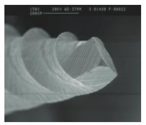
The ProTaper Universal Retreatment (PUR) system was developed specifically for the removal of obturation material during endodontic retreatment. It consists of three instruments: D1 (30.09), D2 (25.08), and D3 (20.07), which were designed to be used in the cervical, middle, and apical thirds of the root canal, respectively. These instruments have a triangular convex section (Figure 1), and only D1 has an active tip to facilitate initial penetration into the filling material.8,9
ProTaper Next is a rotary system made of a NiTi M-wire alloy, which gives greater flexibility and resistance to cyclic fatigue to the instrument. The system includes five shaping instruments: X1 (17.04), X2 (25.06), X3 (30.07), X4 (40.06), and X5 (50.06). All five are characterized by an innovative off-centered rectangular cross section (Figure 2), which has been claimed to give the files a “swaggering” movement as it advances into the root canal.10
ProDesign Logic system (Figure 3), a NiTi alloy with CM heat treatment, increases flexibility and fatigue resistance, has an inactive rounded tip, and variable cutting angle at over the length of the active part, decreasing the screw-in effect. The shaping files are 25.05, 30.05, 35.05, and 40.05.
This study aimed to evaluate, in vitro, the percentage of remaining filling material in mandibular extracted human incisors after removal obturation material with ProTaper Universal Retreatment, ProTaper Next, or ProDesign Logic systems. The null hypothesis is that there is no significant difference in the ability to remove the root canal obturation between the three systems.
Materials and Methods
Teeth selection
The local ethics committee approved this study (NP 1,079,711). Thirty recently extracted human mandibular incisors with complete rhizogenesis, single ovoid canal, the absence of cracks, endodontic treatment, and calcification were selected and stored in 0.1% thymol solution until use. The canal was considered ovoid when the buccal-lingual diameter was twice greater or greater than the mesiodistal diameter in the first two-thirds of the canal measured in the CDR radiographic sensor software.11
The crown was removed by a double-sided diamond disc, standardizing the length of the teeth in 15 mm, confirmed by a digital pachymeter (TESA® DIGIT-CAL® Switzerland). After this procedure, a stainless steel file type K No. 10 (Dentsply Maillefer) was inserted into the root canals until the tip of the instrument was only visible in the apical foramen with the aid of the dental operating microscope (OPMI® Pico; Carl Zeiss Meditec AG, Jena, Germany) under 12.5x magnification. The working length (WL) was determined by subtracting 1 mm from this measurement.


Root canal treatment
The canals were instrumented by a single operator using the NiTi ProTaper Universal rotary files driven to the X-Smart® Plus electric motor (Dentsply Maillefer) at a speed of 300 rpm and 3N. The sequence was performed according to the instructions provided by the manufacturer, using instruments F1, F2, and F3 until the working length was reached. Foraminal patency was maintained with a No. 10 K-file. The root canals were irrigated with 2.5% sodium hypochlorite solution (NaOCl) preceding the use of each instrument during canal shaping, totaling 25 mL of solution per tooth. At the end of the irrigation, the smear layer was removed with 5 mL 17% EDTA for 1 minute, followed by a final flush with 5 mL of 2.5% solution of NaOCl.
The canals were dried with ProTaper F3 absorbent paper points (Dentsply Maillefer) and filled with a ProTaper F3 gutta-percha cone (Dentsply Maillefer) and AH Plus sealer (Dentsply Maillefer) using the Tagger hybrid technique,12 which consists of the lateral obturation technique complemented by the use of the McSpadden gutta condenser. Coronal access was sealed with Coltosol® (Coltene Whaledent, Cuyahoga Falls, Ohio). Buccolingual- and mesiodistal-oriented radiographs were taken with a digital sensor (CDR Schick by Sirona, Dentsply Sirona, Long Island City, New York) to verify the quality of the root canal obturation. Samples were stored in an environment with 100% humidity at 37ºC for 30 days to allow full setting of the sealing. The samples were randomly divided into three groups (n =1 0) using a computerized algorithm (http //www.random.org), according to the techniques of gutta-percha removal.
Removal of filling material
PUR group: The removal of the root canal filling material was performed with a constant speed of 500 rpm for D1 (30.09) and 400 rpm for both D2 (25.08) and D3 (20.07) with a torque of 3Ncm. These instruments were sequentially used in the cervical, middle, and apical thirds, respectively, until reaching WL with the instrument D3. The files were driven on the X-Smart Plus (Dentsply Maillefer) electric motor according to the manufacturer’s instructions.
PN group: In the PN group, X3 file (30.07) was used in the cervical and middle thirds and X2 file (25.06) in the apical third. The instruments were driven in the X-Smart Plus electric motor, in continuous rotation at the speed of 500 rpm and a torque of 3 Ncm. The preparation was performed by root thirds until the instrument reached the WL.
PL group: The ProDesign Logic instrument 30.05 was used in continuous rotation at a speed of 950 rpm and a torque of 4 Ncm.
The instruments were applied in all of the groups using an “in-and-out” motion, and a slight apical pressure was carried out until the instrument reaches the WL followed by a “brushing motion” against the lateral walls during removal file.
Irrigation during filling material removal was performed with 2.5% NaOCl using a total of 25 mL of solution per tooth. Final irrigation was performed with 5 mL 17% EDTA for 1 minute, followed by 5 mL 2.5% NaOCl for each specimen. After the final irrigation, the canals were dried with absorbent points.
Obturation removal was considered complete when there was no obturation material adhered to the instrument used or on the walls of the canal, which were checked with an operating microscope under 12.5x magnification. The instruments were used in three canals and then discarded.13 A single operator with extensive experience in the systems performed the instrumentation of all specimens. No instrument fractures occurred during obturation material removal.
Evaluation of filling material removal
Retreatment time was recorded in minutes for each tooth. The timer was triggered only when the instrument was moving within the canal. The time for irrigation and instrument change was not included.
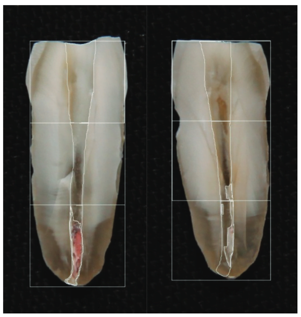
A groove was made on the buccal and lingual surfaces with a double-sided diamond disc (Brasseler USA®) for specimen orientation, and the teeth were cleaved with the aid of a Lecron spatula (Quinelato Company Rio Claro, São Paulo, Brazil). Both halves were photographed using a Sony DSC T20 digital camera (Sony Corporation, Tokyo, Japan) coupled to an operating microscope under 5x magnification. Images were transferred to specific imaging software Image Tool for Windows v.3.00, which was used to design and calculate the areas of the root canal and the remaining filling material, expressed in square pixels. Mean percentage values were then calculated and compared.
The analysis was performed in the cervical, middle, and apical thirds separately, as well as throughout the root canal. Through these measurements, a three simple rule was performed, in which the area of the canal was 100%, and the remaining filling material area equal to x (Figure 4). As shown below:
Remaining area of root filling material X 100 / Canal area = % Remaining area of root filling material
Statistical analysis
The Biostat 4.0 Program performed D’Agostino’s normality test, and the sample presented non-normal behavior. Thus, the Kruskal-Wallis test, supplemented by Student-Newman-Keuls, was used at a significance level of 5% (p <0.05).
Results
The time required for the procedure was 7.17 min for the PUR group, 8.51 min for the PN group, and 5.56 min for the PL group. No statistical significant difference (p> 0.05) was observed among the groups.
The mean obturation material residue was 6.17% in the PUR group, 10.86% in the PN group, and 8.76% in the PL group. No statistical significant difference (p> 0.05) was observed among the groups in the amount of the remaining obturation material, considering the total area of the canal (Table 1).
The descriptive analysis regarding the differences between the three techniques in the cervical, middle, and apical thirds of the roots is presented in Table 2. There was no significant difference between the same thirds of different groups (p> 0.05).In comparisons by thirds within the same sample group, smaller areas of remaining root canal obturation material were found in the cervical third of all groups with significant differences concerning the middle and apical thirds (p <0.05).
Discussion

Due to the advancement of techniques and instruments in endodontics, endodontic treatment has become more predictable and with a high success rate, but failures may still occur.14 The nonsurgical approach has been the preferred treatment to correct such failures because it is the most conservative method and still provides a more favorable long-term result when compared to endodontic surgery.15,16
In endodontic retreatment, the complete removal of the root obturation material is fundamental to allow the effective disinfection of the root canal. However, it is not always possible to completely remove obturation with the currently available methods and instruments.17 Some studies show that there is no significant difference between rotary and manual techniques18,19; others have shown that the manual technique is more efficient20,21; and others have stated that the rotary technique has better performance.22,23 This discrepancy in the results can be attributed to differences in obturation technique, retreatment methods, tooth groups, evaluation methodology, taper, and tip of the instruments.23

Several methods have been used to evaluate root canal cleansing after retreatment, including radiographic evaluation,24 diaphanization,25 longitudinal root cutting with quantitative evaluation of the remnant obturation material,26 qualitative evaluation using a scoring system,27 and micro-CT.28
Although the methodology used in this study is well established in the literature, it has limitations because it provides only two-dimensional pieces of information about a three-dimensional structure,29 and also, the gutta percha can be displaced or lost during the cleavage process.30 Due to these deficiencies, micro-CT has been used because it is a high-resolution, non-destructive method that allows three-dimensional measurement of the volume of the obturation material,29 but the cleavage process remains a viable method.30
In this study, PN and PL rotary systems, which were initially designed for canal instrumentation, were compared by the obturation material remove capacity to the PUR system, which was developed specifically for this purpose. The cervical third presented significantly less remaining obturation material compared to the other thirds of the three groups evaluated.31 In the PUR group, the remaining amount among the thirds was statistically different. In the PN and PL groups, the cervical third had significantly less remaining filling material than the middle and apical thirds, with no statistical difference between them. This result may suggest that the taper of PUR instrument D2 – 25.08 is more adequate compared to PN and PL instruments in the middle third of lower incisors, so that the instrument contacts the walls of the canal more efficiently in this third.
Although the PUR group has the disadvantage that the tip of its last file D3(20.07) is smaller, compared to the other groups that present tip 30(PL) or 25(PN), the favorable result in the removal of the gutta percha is according to the study of Takahashi, et al.2 It can be attributed to the fact that the design of these instruments allows cutting not only the gutta percha, but also a superficial layer of dentin during the retreatment.3
PN and PL systems were as effective in the removal of obturation material as the PUR system. The PN file has a rectangular cross section, and the center of mass and the center of rotation are offset. The device generates a mechanical wave making its movement asymmetrical, which promotes larger areas of contact in the walls of the root canals and, consequently, greater removal of obturation material.32 By the design and high speed with which it is driven, the PL is also capable of removing a large quantity of obturation material. These characteristics may have contributed to the excellent performance of these systems, although they have not been developed for the retreatment procedures.
The results of this study are in part according to the study of Özyürek, et al.,31 who stated that the PN and PUR systems had a similar performance in the removing of the gutta percha. However, concerning the time required for the procedure, the PUR system was faster, differing from our study in which there was no difference between the three groups, as well as the study by Nevares, et al.,32 in which Reciproc and PN did not show differences in the preparation time.
In this study, the three systems evaluated have different alloys — NiTi, M-Wire, and CM-Wire — and did not have significant differences in the removal of filling material, demonstrating that although CM-Wire files (PL group) are more flexible than NiTi files (PUR) and M-Wire files (PN), it had the same ability to remove root canal filling material. It may be suggested that the flexibility of the CM-Wire file assists in the “brushing motion” against the canal walls during the retreatment procedures. This result is in agreement with the study of Rodrigues, et al.,33 where the authors compared the ProDesign R and ProDesign Logic 40.05 (CM-Wire) with Reciproc R 25 (M-Wire) and MTwo 40.04 (NiTi) and had no significant difference in the amount of remaining obturation material.
Conclusion
Based on the results obtained in this in vitro study, it can be concluded that all the evaluated rotary systems were equivalent in removing obturation material from oval canals, but the remaining gutta percha and sealer were observed on the walls of all the canals, regardless of the technique.
Cleaning ovoid canals may be challenging, and in their CE article, Drs. Debelian and Trope address another dilemma in “Cleaning the third dimension.” Click the link to read the article, and subscribers can get 2 credits after passing the quiz!
- Torabinejad, M, Corr, R, Handysides R, Shababang S. Outcomes of nonsurgical retreatment and endodontic surgery: a systematic review. J Endod. 2009;35(7):930-937.
- Takahashi CM, Cunha RS, de Martin AS, et al. In vitro evaluation of the effectiveness of the rotary ProTaper universal retreatment system for gutta-percha removal with or without a solvent. J Endod. 2009;35(11):1580-1583.
- Rios MA, Villela AM, Cunha RS, et al. Efficacy of 2 reciprocating systems compared with a rotary retreatment system for gutta-percha removal. J Endod. 2014;40(4):543-546.
- Mello Junior JE, Cunha RS, Bueno CES, Zuolo ML. Retreatment efficacy of gutta-percha removal using the microscope clinical and ultrasonic instruments: part I — an ex vivo study. Oral Surg Oral Med Oral Pathol Oral Radiol Endod. 2009;108(1):59-62.
- Bueno CES, Delboni MG, Araujo RA, Carrara HJ, Cunha RS. Effectiveness of rotary and hand files in gutta-percha and sealer removal using chloroform or chlorhexidine gel. Braz Dent J. 2006;179(2):139-43.
- de Carvalho Maciel AC, Zaccaro Scelza MF. Efficacy of automated versus hand instrumentation during root canal retreatment: an ex vivo study. Int Endod J. 2006;39(10):779-784.
- Baratto Filho F, Ferreira EL, Fariniuk LF. Efficiency of the 0.04 taper ProFile during the re-treatment of gutta-percha-filled root canals. Int Endod J. 2002;35(8):651-654.
- Marfisi K, Mercade M, Plotino G, et al. Efficacy of three different rotary files to remove gutta-percha and Resilon from root canals. Int Endod J. 2010;43(11)1022-1028.
- Hussne RP, Braga LC, Berbert FL, Buono VT, Bahia MG. Flexibility and torsional resistance of three nickel-titanium retreatment instrument systems. Int Endod J. 2011;44(8):731-738.
- da Silva Limoeiro AG, Dos Santos AH, De Martin AS, et al. Micro-computed tomographic evaluation of 2 nickel-titanium instrument systems in shaping root canals. J Endod. 2016;42(3):496-499.
- Martins MP, Duarte MAH, Cavenago BC, Kato AS, Bueno CES. Effectiveness of the ProTaper Next and Reciproc systems in removing root canal filling material with sonic or ultrasonic irrigation: a micro-computed tomographic study. J Endod. 2017;43(3):467-471.
- Tagger M, Tamse A, Katz A, Korzen BH. Evaluation of the apical seal produced by a hybrid root canal filling method, combining lateral condensation and thermatic compaction. J Endod. 1984;10(7):299-303.
- Rödig T, Kupis J Konietschke F, et al. Comparison of hand and rotary instrumentation for removing gutta-percha from previously treated curved root canals: a microcomputed tomography study. Int Endod J. 2014;47(2):173-182.
- Siqueira JF Jr. Aetiology of root canal treatment failure: why well-treated teeth can fail. Int Endod J. 2001;34(1):1-10.
- Hammad M, Qualtrough A, Silikas N. Three-dimensional evaluation of the effectiveness of hand and rotary instrumentation for retreatment of canals filled with different materials. J Endod. 2008;34(11):1370-1373.
- Só MVR, Saran C, Magro ML Vier-Pelisser FV, Munhoz M. Efficacy of ProTaper retreatment system in root canals filled with gutta-percha and two endodontic sealers. J Endod. 2008;34(10):1223-1225.
- Ersev H, Yilmaz B O, Dinçol ME Daglaroglu R. The efficacy of retreatment ProTaper Universal rotary single instrumentation to remove gutta-percha cones cemented with several endodontic sealers. Int Endod J. 2012;5(8):756-762.
- Barrieshi-Nusair KM. Gutta-percha retreatment: effectiveness of nickel-titanium rotary instruments versus stainless steel hand files. J Endod. 2002;28(6):454-456.
- Kfir A, Tsesis I, Yakirevich E, Matalon S, Abramovitz I. The efficacy of five techniques for removing root material: microscopic versus radiographic evaluation. Int Endod J. 2012;45(1):35-41.
- Zmener O, Pameijer CH, Banegas G. Retreatment efficacy of hand versus automated instrumentation in oval-shaped root canals: an ex vivo study. Int Endod J. 2006;39(7):521-526.
- Xu L, Zhang L, Zhou X, et al. Residual filling material in dentinal tubules after gutta-percha removal observed with scanning electron microscopy. J Endod. 2012; 38(3):293-296.
- Bernardes RA, Duarte MAH, Vivan RR, et al. Comparison of three retreatment techniques with ultrasonic activation in flattened canals using micro-computed tomography and scanning electron microscopy. Int Endod J. 2016;49(9):890-897.
- Saad AY, Al-Hadlaq SM, Al-Katheeri NH. Efficacy of two rotary NiTi instruments in the removal of gutta-percha during root canal retreatment. J Endod. 2007;33(1):38-41.
- Masiero AV, Barletta FB. Effectiveness of different techniques for removing gutta-percha during retreatment. Int Endod J. 2005;38(1):2-7.
- Schirrmeister JF, Wrbas KT, Meyer KM, Altenburger MJ, Hellwig E. Efficacy of different rotary instruments for gutta-percha removal in root canal retreatment. J Endod. 2006;32(5):469-472.
- Imura N, Kato AS, Hata GI, et al. A comparison of the relative efficacies of four hand and rotary instrumentation techniques during endodontic retreatment. Int Endod J. 2000;33(4):361-366.
- Hülsmann M, Bluhm V. Efficacy, safety and cleaning ability of different rotary NiTi instruments in root canal retreatment. Int Endod J. 2004;37(7):468-476.
- Fruchi LC, Ordinola-Zapata R, Cavenago BC, et al. Efficacy of reciprocating filling instrument for removing the material in curved canals obturated with a single-cone technique: micro-computed tomographic analysis. J Endod. 2014;40(7):1000-1004.
- Zuolo AS, Zuolo ML, da Silveira Bueno CE, Chu R, Cunha RS. Evaluation of the efficacy of TRUShape and Reciproc file systems in the removal of root filling material: an ex vivo micro-computed tomographic study. J Endod. 2016;42(2):315-319.
- Gu LS, Ling JQ, Wei X, Huang XY. Efficacy of ProTaper Universal rotary retreatment system for gutta-percha removal from root canals. Int Endod J. 2008;41(4):288-295.
- Özyürek T, Demiryürek EÖ. Efficacy of different nickel-titanium instruments in gutta-percha removal during root canal retreatment. J Endod. 2016;42(4):646-649.
- Nevares G, de Albuquerque DS, LG Freire, et al. Efficacy of ProTaper NEXT compared to Reciproc in obturation material from severely curved root canals: a micro-computed tomography study. J Endod. 2016;42(5):803-808.
- Rodrigues CT, Duarte MA, de Almeida MM, de Andrade FB, Bernardineli N. Efficacy of CM-Wire, M-Wire, and Nickel-Titanium instruments for removing filling material from curved root canals: a micro-computed tomography study. J Endod. 2016;42(11):1651-1655
Stay Relevant With Endodontic Practice US
Join our email list for CE courses and webinars, articles and more..
