Influence on Fees, Reputation, Longevity, and Because: Part 10 of Anatomy Matters

Dr. John West discusses four more reasons why anatomy should matter

Introduction

After nine installments of Anatomy Matters,1-9 I am beginning to feel like a broken record. So, I am writing Anatomy Matters, Part 10, by taking a different perspective — rather than continuing to write about endodontic patient treatments that fail due to underfilled root canal systems, my objective is to pique your interest in a new way, by telling you that the future standard form of procedure reimbursement will be how well endodontists can prove they have filled the cleaned root canal system. It will be called “Pay for Performance” (PFP). Check out QForma on the Internet and begin to be educated that we are being graded; we are being measured; we are being watched.

Figures 1A and 1B: Anatomy and Fees. The greater the endodontic value, the greater the clinician’s value and therefore, the higher the set fee. 1A. Posttreatment perpendicular image of recent endodontic finish of maxillary left second molar. Meanwhile, maxillary left first molar had been previously treated and operator origin unknown. First molar has internal POE transportation of four canals and therefore no sealed POEs (violation of mechanical objective No. 4). Second molar has three canals and more than twice as many visibly obturated POEs. The future Pay for Performance reimbursement method will place more value on the second molar two-dimensional endodontic result than the first molar. 1B. Oblique image

I have divided my article into four sections: 1) Anatomy and fees, 2) Anatomy and reputation, 3) Anatomy and longevity and 4) Anatomy and because.

Four proofs that anatomy matters

1. Anatomy and fees

Figures 2A-2E: Anatomy and reputation. Are you the “go to” endodontic resource when the treatment is challenging, or the success of the result is critical? 2A. Pretreatment image of symptomatic maxillary second premolar with two strikes against it: 1) Nonsurgical and surgical Endodontic Seal already attempted. 2) Compromised crown/root ratio and recently restored with post, foundation, and crown. Most dentists would treatment plan removal and replacement with implant, but since not fractured, symptoms were simply due to undersealed root canal system. Skilled nonsurgical disassembly and Endodontic Seal attempt should be in the patient’s best biologic and financial interest. Do you have the confidence, the skill, and the reputation to realistically offer nonsurgical endodontic option? 2B. Image of post removed. 2C. Post removed. 2D. Posttreatment image demonstrating significant distal POE sealed. 2E. Eight-year posttreatment image demonstrating rejuvenation of the lamina dura and periodontal ligament. The original restorative has structurally remained intact and the tooth is functioning normally

What this means is that some day (sooner than we think), insurance companies, government, and the consumer (our patients), will know the following:

  • how good dentists’ crown margins are
  • the rate of their veneer success/failure
  • the number of healthy teeth in the dentists’ patient population
  • if their root canal obturations are solid
  • if their endodontic preps have continuously tapering funnel shapes
  • how clean the clinicians’ root canal systems are
  • how many visible portals of exit (POEs) they fill per shaped canal

These will be the quantified measurements that will determine our value and, therefore, our fees (Figure 1).

Forget that anatomy matters to some and not others. I do know this, that endodontists want more patients. And if endodontists are truly going to be paid on performance, and the pretreatment and posttreatment radiographic images are our only metric, then biologic success is not the only reason to make the perfect access cavity, to remove all dentin triangles, to prepare a glide path to the canal terminus no matter what, and to produce appropriate shapes that protect the ferrule and yet are sufficient for gutta percha and sealer fluid hydraulics for 3D obturation.

You may say that fee-for-service is never going to be based on the radiographic quality of my obturated properly shaped root canal system, let alone the number of POEs visibly sealed on a periapical radiograph. Who’s counting? Maybe we should take a cone-beam computed tomography (CBCT) 3D posttreatment image, too, in order to grade and pay for our technical result. I believe that your referring dentist is counting and that your reputation is counting.

2. Anatomy and reputation
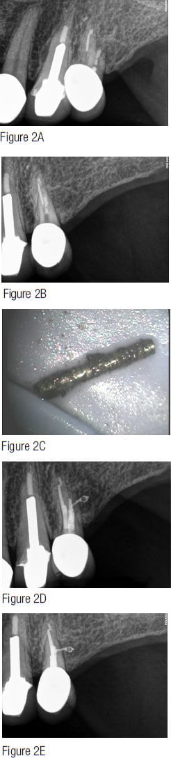
Realistically, I don’t think any PFP future is near in time, but the quality of our measured technical result (the radiographs or digital imaging) does influence the endodontist’s reputation and, quite frankly, our “busy-ness” (Figure 2). For example, if an endodontist cannot predictably perform the big three “F’s” — find all the canals, follow all the canals to their termini, and finish the canals (smooth, funnel shape, 3D clean, conefit, and 3D obturation, or at least the appearance of 3D obturation) — then the word gets out pretty fast, and that endodontist suddenly finds himself or herself competing for referrals not in the top 10% of the market but the bottom 10%. The bottom 10% seems willing to treat for the lowest fee to get some referrals. Often the bottom 10% number of referrals is insufficient for significant profitability, and these patients often lack dental value. Either way, competing in the bottom 10% is dissatisfying, de-energizing, and physically and emotionally exhausting. At least for me, when I go faster or base my treatment on time, I begin to risk making mistakes. I block; I tear; I break things. None of these outcomes is good for the reputation.

3. Anatomy and longevity
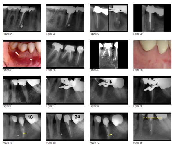
It’s time for me to get off my “strive for quality” soapbox. Let’s just surmise that I have not made a good argument: Our fees will never be based on a radiographic image, and referring dentists don’t care about the only measure of quality, aside from patient feedback, available — the X-ray or image that the endodontist sends back to the dentist after endodontic finish. Is there any other reason to do our best and operate as if anatomy matters? Yes, the better that root canal systems are obturated, the longer they last, and the better the patient investment in saving endodontically diseased teeth.10 And since our population is aging, each new decade requests that dentistry, including endodontics, must last a longer time. It must be built to last (Figure 3).11

4. Anatomy and because
The following text is an excerpt from John F. Kennedy’s “Moon Speech” at Rice University on September 12, 1962:

We choose to go to the moon. We choose to go to the moon in this decade and do the other things, not because they are easy, but because they are hard, because that goal will serve to organize and measure the best of our energies and skills, because that challenge is one that we are willing to accept, one we are unwilling to postpone, and one which we intend to win. … Many years ago the great British explorer George Mallory, who was to die on Mount Everest, was asked why did he want to climb it. He said, “Because it is there.”

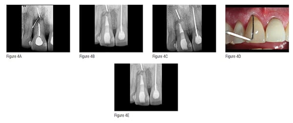
“Because it is there.” Well, consistently performing great endodontics is here; and while great endodontics has never been rocket science, it has always taken a certain willingness, and it has always, like the race to the moon, served to organize and measure “the best of our energies and skills” (Figure 4). It may be that the anatomy doesn’t matter. The LEO may not develop, and the LEO may heal regardless of the quality of the Endodontic Seal.

Instead, it is the “Because” that is the because.

Figures 3A-3P: Anatomy and longevity. Can you tell your patient that your endodontic treatment can predictably last them the rest of their lives? 3A. Posttreatment image of mandibular right second premolar classic nonsurgical warm gutta-percha technique obturation. 3B. 32-year posttreament image. 3C. Pretreatment and downpack image of mandibular left FPD abutment. 3D. 29-year posttreatment image. 3E. Gutta-percha traced sinus tracts from mandibular molar and premolar of previous nonsurgical followed by surgical underfilled root canal systems. 3F. Pretreatment image. 3G. 27-year posttreatment image. 3H. 27-year posttreatment clinical with no sinus tracts. 3I. Pretreatment of mandibular left second premolar with lateral LEO. 3J. First instrument to radiographic terminus. 3K. Instrument following in and through distal lateral POE. 3L. Conefit. 3M. 18-month posttreatment with arrow pointing to lateral POE position. 3N. 24-month posttreatment revealing increase in lateral LEO size increase. 3O. Post-surgical retreatment image of amalgam surgically sealing lateral POE (arrow). 3P. 34-year posttreatment image
Figures 4A-4E: Anatomy and because. Sometimes we do something because it is there; because we want to “organize and measure the best of our ‘energies and skills.’” 4A. Pretreatment image of seemingly hopeless tooth with gutta-percha cone tracing sinus tract to resorbed root end with sectioned silver cone. 4B. “Because it is there and possible,” patient elected to attempt nonsurgical endodontic retreatment. He did not want an implant. Image shows pack film. 4C. Same pack film placed more apically in order to show there was no apparent radiolucency around section silver cone in apical root remnant, and treatment plan was to leave the root tip unless future evidence of pathology. 4D. Two- year posttreatment with healthy gingival probing and no sinus tract. Patient has elected to proceed with connective tissue graft in order to improve gingival levels. 4E. Two-year posttreatment image with healthy attachment apparatus. Retreatment of adjacent maxillary central is scheduled with safe “walking” bleach intended to improve gingival root discoloration

Conclusion

For the purposes of this 10th installment of Anatomy Matters, I have suggested that there are at least four reasons to consider doing better endodontics and that anatomy does matter: 1) value, 2) reputation, 3) longevity, and 4) because. I would offer that for most of us, we can do better endodontics by simply slowing down, improving technical skills, or simply being more intentional. I believe any of us can do as much as we want. Start with the easy cases, and then move to the more complicated. While some people intend on having you do endodontics at a lower, lower, lower level, I guess I am intent on having you do endodontics at a higher, higher, higher level. And I have enough experience of not only doing endodontics but also teaching it that I am right, and it can be done. Professor Herb Schilder once said, “It’s not hard to be the best. There’s no competition.” Compete in the top 10%; there is less competition there. But really, to me, this means compete in the top 10% of your own personal potential. Being and doing your best in delivering endodontic excellence has nothing to do with measuring how good I am compared to you. It has to do with how good I am compared to me.

John West, DDS, MSD, the founder and director of the Center for Endodontics, British-born Dr. John West continues to be recognized as one of world’s premier educators in clinical and interdisciplinary endodontics. John West received his DDS from the University of Washington in 1971 where he is an Affiliate Associate Professor. He then earned his MSD in endodontics at Boston University Henry M. Goldman School of Dental Medicine in 1975 where he is a clinical instructor and has been awarded the Distinguished Alumni Award. Dr. West has presented unmatched endodontic continuing education in North America, South America, and Europe while maintaining a private practice in Tacoma, Washington. Dr. West is a clinical visionary, an inventor, a teacher, and a coach for any dentist who wants to experience the possibilities of endodontics in his/her practice. He coauthored Obturation of the Radicular Space with Dr. John Ingle in Ingle’s 1994 and 2002 editions of Endodontics and was senior author of Cleaning and Shaping the Root Canal System in Cohen and Burns 1994 and 1998 Pathways of the Pulp. He has authored Endodontic Predictability in Dr. Michael Cohen’s 2008 Quintessence text Interdisciplinary Treatment Planning: Principles, Design, Implementation, and Michael Cohen’s 2010 Quintessence text Interdisciplinary Treatment Planning Volume II: Comprehensive Case Studies and is lead author of Esthetic Management of Endodontically Treated Teeth in Ronald Goldstein’s “in print” third edition of Esthetics in Dentistry. Dr. West’s memberships include 2009 president and fellow of the American Academy of Esthetic Dentistry and 2010 president of the Academy of Microscope Enhanced Dentistry, the Northwest Network for Dental Excellence, and the International College of Dentists. He is a 2010 consultant for the ADA’s prestigious ADA Board of Trustees where he serves as a consultant to the ADA Council on Dental Practice. Dr. West further serves on the Henry M. Goldman School of Dental Medicine’s Boston University Alumni Board. He is a Thought Leader for Kodak Digital Dental Systems and serves on the editorial advisory boards for: The Journal of Esthetic and Restorative Dentistry, Practical Procedures and Aesthetic Dentistry, and The Journal of Microscope Enhanced Dentistry.

Web          www.centerforendodontics.com

E-mail       johnwest@centerforendodontics.com

Phone       1-800-900-7668 (ROOT)

Fax            253-473-6328

  1. West J. Anatomy matters. Endodontic Practice US. 2012;5(2):14-16.
  1. West J. Anatomy matters — part 2. Endodontic Practice US. 2012;5(4):26-27.
  1. West J. Anatomy matters part 3. Furcal endodontic seal heals furcal lesion of endodontic origin. Endodontic Practice US. 2012;5(6):22-24.
  1. West J. Anatomy matters. Long-term case report. Endodontic Practice US. 2013;6(1):50-51.
  1. West J. Anatomy matters. Root canal system anatomy only matters when it matters. Endodontic Practice US. 2013;6(2):56-58.
  1. West J. Anatomy matters. Do lateral canals really matter? Part 6. Endodontic Practice US. 2013;6(3):52-53
  1. West J. Anatomy matters. “What’s it all about?” Part 7. Endodontic Practice US. 2013;6(4):52-54.
  1. West J. Anatomy matters. “Could it all simply be a coincidence?” Part 8. Endodontic Practice US. 2013;6(5):52-55.
  1. West J. Anatomy matters. Endodontic accountability: The “X” factor, part 9. Endodontic Practice US. 2014;7(1):43-47.
  1. West JD. Implants versus endodontics: “As the pendulum swings.” Dent Today. 2014;33(1):10-12.
  1. Kurzweil R. The Singularity is Near: When Humans Transcend Biology. New York, NY: Penguin; 2005.

Stay Relevant With Endodontic Practice US

Join our email list for CE courses and webinars, articles and more..

Scroll to Top