Kerr ZenFlex One files were integral in helping Dr. David King treat this painful and non-restorable tooth.
Dr. David King treats a tooth that was interfering with quality of life
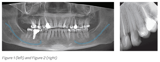
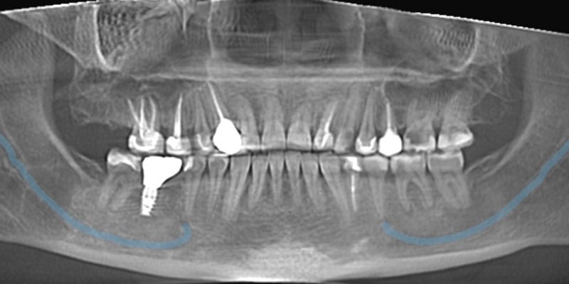
The patient presented for examination of pain that had persisted over the previous few days in the upper left (UL) area, specifically tooth No. 11. The patient described that the pain was moderate and interfering with certain aspects of life. Radiographs were taken to evaluate for carious and non-carious pathology. After examining the radiographs, it was determined that tooth No.14 was non-restorable, and tooth No. 11 needed endodontic treatment, a core, and crown to restore health (Figures 1 and 2).
After discussing all treatment options, the patient opted for endodontic therapy and
buildup. Nitrous oxide was declined, so not used. Treatment began with local anesthetic that included: 1.7cc (1 carpule) 4% Articaine HCl w/1:100k epi. infiltration.
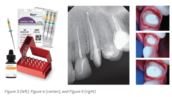
Further examination noted that decay had progressed into the nerve. The decay was removed and the canal accessed, after which the canal was cleaned and shaped with Kerr ZenFlex™ ONE (Figure 3).
After cleaning and shaping, the canal, which was determined to be 23.5 mm in length,was rinsed, irrigated, and agitated with 3ccs each of NaOCl and EDTA. The canal was sized and shaped with a ZenFlex™ ONE Medium (Green 35.06/25 mm) file to working length using crown-down technique and substantial irrigation. The anatomical location, near the anatomic apex, was slightly distally angulated.
The canal was dried, BC sealer was placed, and the canal was obturated with warm condensation. Then, the space was prepared for a FibreKleer™ 4x Tapered 1.5 mm post(Figure 3). The post was put in place and bonded with Optibond™ Universal (Figure 4).
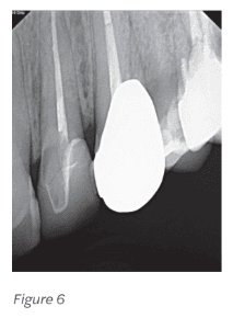
The patient had opted in for a crown because the caries/failing restoration had extended to greater than two-thirds of the occlusal table, and endodontic treatment had just been completed. The tooth was reduced, and occlusion clearance and margin height were checked (Figure 5). The final crown, in shade A2, was milled in house and seated same day (Figure 6). The final crown was cemented with Optibond™ Universal bond and Maxcem Elite™ Cement.
This case study was provided by Kerr.
Read more about how Kerr Dental is addressing the diverse needs of endodontic practices here: https://endopracticeus.com/kerr-dental/
Stay Relevant With Endodontic Practice US
Join our email list for CE courses and webinars, articles and more..
