Drs. Jorge Alberdi and Fernando Goldberg compare canal transportation in simulated double-curved root canals
Summary
Objective
To compare canal transportation in simulated double-curved root canals after instrumentation with the Reciproc® R25 and Reciproc® Blue R25 files.
Materials and methods
Thirty Endo Training Block-S models were dyed with black India ink and photographed. The resin blocks were randomly divided into two groups: A and B (n = 15). The canals were instrumented with the Reciproc R25 (Group A) and Reciproc Blue R25 (Group B) systems. After instrumentation, the canals were dyed again and photographed under the same conditions. The pre-instrumentation images were superimposed on the post-instrumentation images. A measurement template, divided into 16 equal cells of 1 mm, was placed on each instrumentation in each canal of the 30 blocks. The visual evaluation was conducted in cells 1 to 10 according to the following categorization: 0 = centered and 1 = transported. Apical transportation was evaluated for each group. The position of the foramen after instrumentation was classified into three categories: 0 = centered, 1 = transported, and 2 = perforated. Groups were compared for the variable “modification” within each cell by the chi-squared test or Fisher’s exact test, as appropriate, and for the variable “foramen” by the chi-squared test. In all cases, the level of significance was P < 0.05.
Results
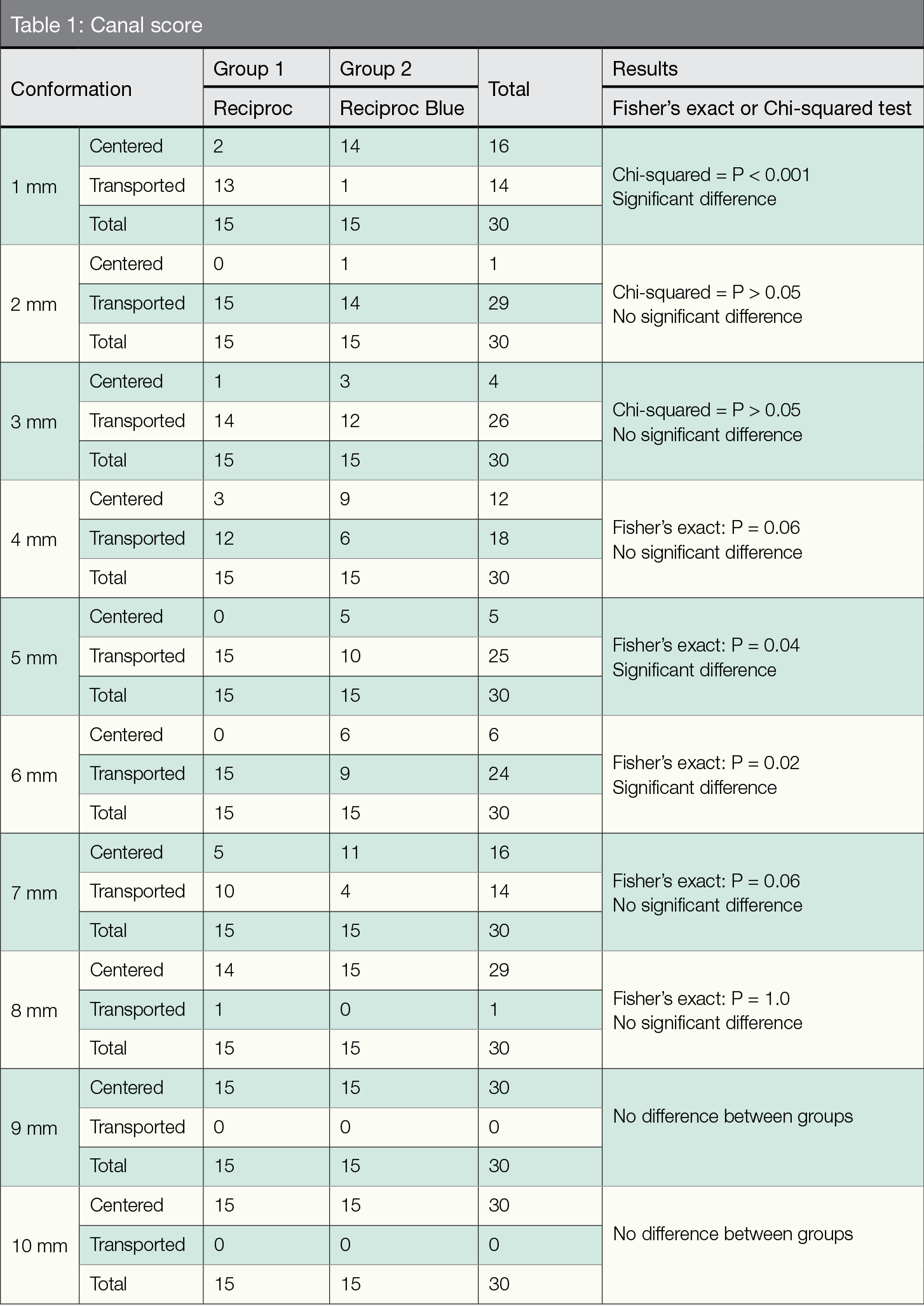
The differences between groups were significant at 1 mm, 5 mm, and 6 mm but not significant at 2 mm, 3 mm, 4 mm, 7 mm, 8 mm, 9 mm, or 10 mm. With regard to apical transportation, the difference between groups was significant (P < 0.001).
Conclusion
Compared with the Reciproc R25 file, the Reciproc Blue R25 file produced a more centered canal that more accurately represented the original anatomy of the simulated double-curved root canal.
Objective
The objective of this study was to compare, through the superposition of photographic images, canal transportation in simulated double-curved root canals (Endo Training Block-S) after instrumentation with Reciproc R25 and Reciproc Blue R25 files.
Introduction
Nickel-titanium (NiTi) instruments have been and continue to be widely used for the preparation of root canals. The great advantage of these instruments is their flexibility, which allows the technique for preparing curved canals to become more predictable. However, in these situations, there is a risk of torsional fracture and/or cyclical fatigue, which compromises the prognosis of the endodontic treatment. Different alloys and instrument sections have been proposed to increase flexibility and resistance to fatigue.1
The thermal treatment of NiTi alloys has been an important advance in improving the mechanical properties of the instrument, resulting in a better centered and safer preparation of the root canal.2-3 Manufacturers have introduced several heat-treated NiTi alloys, such as control memory wire (Coltène Whaledent, Cuyahoga Falls, Ohio), M-Wire™ (Dentsply Tulsa Dental Specialties, Tulsa, Oklahoma), and R-phase wire (SybronEndo, Orange, California).
A new generation of instruments (Vortex Blue® and ProTaper Gold® rotary files (Dentsply Sirona Endodontics, Tulsa, Oklahoma) are subjected to a complex heating-cooling treatment that results in a visible layer of titanium oxide on the surface of the instrument. This treatment controls the transition temperatures, creating a shape-memory alloy that the manufacturer claims improves the mechanical and performance properties of NiTi instruments. Also on the market is the HyFlex™ rotary system (Coltène Whaledent), with an alloy of similar characteristics and other control memory products.4
In addition to thermal treatment, a new kinematics, the reciprocating motion, extends the useful life of NiTi instruments and their resistance to fatigue compared with continuous rotation. Reciprocating instruments travel a shorter angular distance than rotary instruments, so they are subject to lower tension values and prolonged fatigue life.5 Reciproc (VDW, Munich, Germany) and WaveOne® (Dentsply Sirona, Baillagues, Switzerland) are the main examples of systems available for the preparation of the root canals using reciprocating motion; both instruments are currently manufactured with NiTi M-wire, a heat-treated alloy marketed as Blue and Gold versions, respectively. Thermal treatment when NiTi is in the crystalline phase enables very good flexibility and little elastic memory, which prevents the instrument from recovering its shape inside the root canal, conforming better to the anatomy of the canal. In addition to thermal treatment, the WaveOne Gold® (Dentsply Sirona) has an updated cross-sectional instrument shape: a parallelogram.6 The design of the Reciproc Blue (VDW) remains exactly the same as its M-wire version; the only difference between the two Reciproc versions is the heat treatment used during their manufacture.
Numerous authors have used resin models with double-curved simulated canals (DCSCs) with the purpose of evaluating the conformations generated by different mechanized instrumentation systems.1-6 These models are marketed with different curvatures that resemble root canals with difficult anatomies. DCSC models are manufactured with similar morphology, working length, caliber, and angle of curvature to real canal anatomy, which allows the performance of different endodontic preparation systems to be compared without having to manage the anatomical variables of human teeth.
In the present study, the conformations produced by the Reciproc R25 and Reciproc Blue R25 files in simulated double-curved root canals were compared to determine whether the alloy treatment modifies their performance in keeping the root canal centered.
Materials and methods
Simulated canals
In the present study, 30 Endo Training Block-S resin models (Dentsply Sirona) with these features were used:
- simulated canals of double curvature (S-shaped)
- a conicity of 0.02
- an apical diameter of 0.15 mm
- a length of 16 mm
The respective angles and radii were 30 mm and 5 mm for the coronal curvature and 20 and 4.5 mm for the apical curvature. The permeability of the canals was confirmed by passing a K No.10 file (Dentsply Sirona) beyond the foramen. Next, all the DCSCs were dyed with black India ink (Pelikan, Argentina) and injected with a Max-i-Probe 30-G needle (Dentsply-Rinn, Elgin, Illinois). The stained DCSCs were photographed using a cell phone digital camera (iPhone® 6S plus, 12 megapixels) on a fixed support and under the same image output parameters. Subsequently, the resin blocks were randomly divided into two groups, A and B (n = 15 canals/group), and numbered 1 to 15 in each group.
S-shaped canal instrumentation
The DCSCs were instrumented using the Reciproc R25 and Reciproc Blue R25 systems according to the manufacturer’s instructions and irrigated with 2 ml of distilled water using a Max-i-Probe® 30G needle (Dentsply-Rinn, Elgin, USA) before, during, and after DCSC instrumentation.
Groups
Group A: Fifteen DCSCs were instrumented with the Reciproc R25 file (VDW), apical caliber No. 25 and conicity 0.08, operated in the “RECIPROC” mode of the X-Smart Plus motor (Dentsply Sirona). The instruments were used with a pecking motion, advancing approximately 3 mm, according to the manufacturer’s instructions. The whorls of the instrument were cleaned with alcohol-moistened gauze before each new entry.
Group B: Fifteen DCSCs were instrumented with the Reciproc Blue R25 file (VDW), apical caliber No. 25 and conicity 0.08. The instrumentation was performed with the same motor and procedure as group A.
Once the instrumentation was finished, the DCSCs were stained again following the same procedure, and the resin blocks were photographed again under the same conditions.
Image analysis and evaluation of the preparation of the canal
The pre- and post-instrumentation images of each sample were copied and pasted into a presentation slide of Keynote software (Apple Inc., Cupertino, California). The preoperative black image of the DCSC was converted into a pink image using Keynote®. Then the pre-instrumentation image was superimposed with the post-instrumentation image. A measurement template, prepared with the same software and divided into 16 equal cells representing 1 mm each, was placed from the apical region to the coronal portion of the canal on each of the DCSCs of the 30 resin blocks.
Visual evaluations were conducted in cells 1 to 10 according to the following categorization: 0 = centered, 1 = transported. Cells 1 to 3 corresponded to 3 mm of the apical curve, 3 to 6 to 3 mm of the coronal curve, and 7 to 10 to 3 mm of the straight portion of the DCSC (Figure 1). The results were entered in a score table for each sample. The apical transportation for groups A and B was evaluated according to tables prepared for that purpose. The position of the foramen after instrumentation was classified into three categories: 0 = centered, 1 = transported, and 2 = perforated. These data were entered into the table. The variable “modification” within each cell was compared using the chi-squared test or Fisher’s exact test, as appropriate. The variable “foramen” was compared between groups using the chi-squared test. In all cases, the level of significance was P < 0.05.
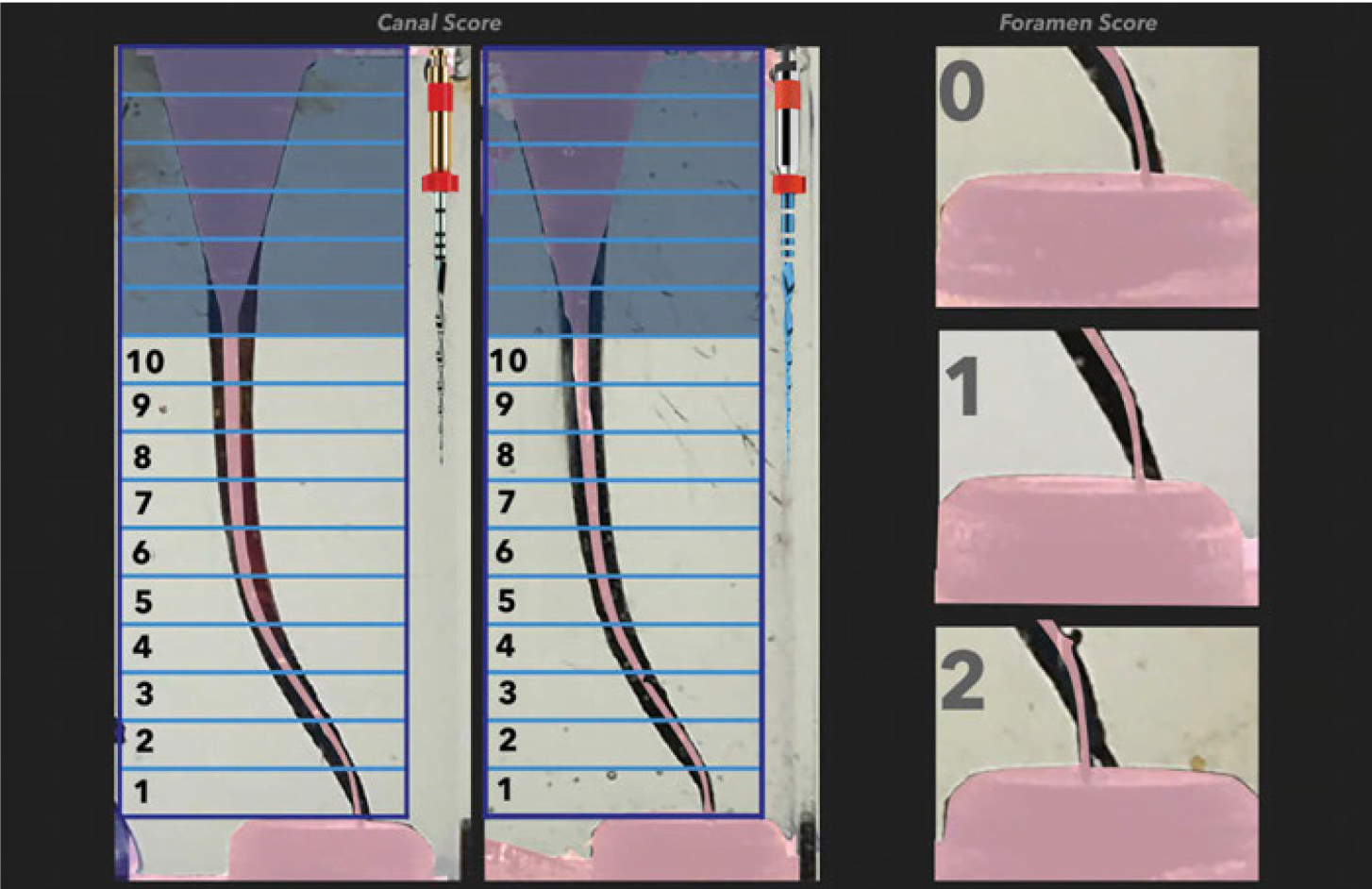
Figure 1

Chi-squared = 17.4; P < 0.001 (significant differences between systems)
Results
The differences between groups were significant at 1 mm, 5 mm, and 6 mm and not significant at 2 mm, 3 mm, 4 mm, 7 mm, 8 mm, 9 mm, or 10 mm (Table 1). With regard to apical transportation, the significant difference between groups was significant (P < 0.001) (Table 2).
Discussion
In this study, resin blocks with simulated double-curved root canals were used to reproduce clinical situations with complicated anatomies. The use of the Endo Training Blocks (Dentsply Sirona) enables a more realistic comparison of the conformation produced by the different instrumentation systems. Their use makes standardization of the experimental conditions possible.8,9 The resin blocks can be photographed, measured, and evaluated before and after the canal-shaping procedure.4 Care must be taken when extrapolating the experimental results of simulated canals to clinical applications because there is a difference in hardness between resin and dentin.7 In addition, the dental elements of actual root canals have numerous infractuosities and varying cross sections along their anatomy from coronary to apical, while the canals of the resin blocks have a circular cross-section throughout the entire length of the path, without irregularities.
De Deus, et al.,10 found that the instruments manufactured with the NiTi Blue alloy were more flexible than those manufactured with NiTi M-wire. Gao, et al.,11 obtained similar results when comparing Vortex rotary files (Dentsply Tulsa Dental Specialties) manufactured with both alloys. Gagliardi, et al.,12 compared ProTaper Universal® (Dentsply Maillefer), ProTaper Next® (Dentsply Maillefer), and ProTaper Gold (Dentsply Maillefer) and found that both ProTaper Gold and ProTaper Next showed less transportation of the root canal. ProTaper Universal and ProTaper Gold have the same geometric design but are made from different alloys. The greater flexibility of the ProTaper Gold alloy, combined with the decreased recovery strength of the instrument, could explain why the root canals remained better centered during instrumentation.12,13 Özyürek, et al.,6 highlighted that files made with alloys of greater flexibility were better centered in simulated S-shaped canal preparations. Similar results were obtained in the present study when comparing two identically designed files of different alloys. The NiTi Blue showed, as did the NiTi Gold alloy, a loss of elasticity resulting in a weaker recovery force. Along these lines, Pongione, et al.,14 noted the benefit of using more flexible instruments in curved canals to reduce the tendency for iatrogenic errors and to facilitate apical preparations while maintaining the original morphology of the root canal.
Yared15 proposed the possibility of using a single file for root canals in 2008, for which he used a reciprocating-motion Universal ProTaper F2 file (Dentsply Maillefer). Subsequently, the Reciproc file was introduced into the market and has been widely studied. Zuolo, et al.,16 found in vivo that the Reciproc R25 file (VDW) was 32% more effective than the C-Pilot manual instruments No. 06, No. 08, and No. 10 (VDW) in permeabilizing and negotiating the MB2 canal of upper molars.
Plotino, et al.,17 reported the high cutting efficiency of the Reciproc instrument (VDW) compared with the WaveOne file and emphasized that its cross-sectional design appeared to be the determining factor of its cutting capacity. Recently, Topcuoglu and Topcuoglu18 revealed that the Reciproc Blue R25 and R40 (VDW) files had greater resistance to cyclic fatigue than their predecessors Reciproc R25 and R40 (VDW), manufactured with NiTi M-wire, when they instrumented S-shaped artificial canals. Keskin, et al.,19 described similar results, observing that the Reciproc Blue R25 files (VDW) had higher cyclic fatigue resistance than the WaveOne Gold Primary and Reciproc R25 files.
In the present study, the Reciproc R25 and Reciproc Blue R25 files were used, taking into account that the S-shaped curvature of the simulated canals required using instruments of smaller caliber and conicity.20
Conclusion
Under the conditions of this study, the Reciproc Blue R25 file produced a more centered canal that more accurately represented the original anatomy of the double-curvature simulated canal than did the Reciproc R25 file.
Acknowledgments
Appreciation to Dr. Ricardo L. Macchi for his collaboration in the statistical evaluation.
 Jorge Alberdi, DDS, is a specialist in endodontics, and auxiliary professor at the Endodontic Department of the Dental School at Universidad del Salvador/Asociación Odontológica Argentina (USAL/AOA) in Buenos Aires, Argentina. He is Director of the Postgraduate Endodontic Course at Círculo Odontológico de Rosario (COR), Rosario, Argentina, and is in private practice in endodontics and restorative post-endodontics in Las Rosas, Santa Fe, Argentina. Dr. Alberdi is also director of the Operative Microscope in Endodontics postgraduate course at the Institute Troiano Dentistry, Rosario, Argentina.
 Fernando Goldberg, DDS, PhD, is Professor Emeritus of the Endodontic Department of the Dental School at the Universidad del Salvador- Asociación Odontológica Argentina. He is also a professor at the Department of Endodontics at the Dental School, Universidad de Buenos Aires, Argentina. Dr. Goldberg is a Life Member of the American Association of Endodontists. He is author of the book Materiales y Técnicas de Obturación Endodóntica. Ed. Mundi. 1982 and editor of Endodoncia Técnica y Fundamentos, Ed. Med. Panamericana, Buenos Aires, Argentina, 2002 and 2nd edition 2012. He has also had numerous papers published in local and international journals.
Disclosure: The authors declare that there are no conflicts of interest related to this study.
- Zmener O, Pameijer CH, Álvarez Serrano S. Análisis histométrico de la capacidad de dos sistemas mecanizados para la instrumentación y conformación de conductos curvos simulados. Rev Asoc Odontol Argent. 2011;99:325-33.
- Berutti E, Cantatore G, Castellucci A, et al. Use of nickel-titanium rotary Pathfile to create the glide path: comparison with manual preflaring in simulated root canals. J Endod. 2009;35:408-412.
- Berutti E, Paolino DS, et al. Root canal anatomy preservation of WaveOne reciprocating files with or without glide path. J Endod. 2012;38:101-104.
- Shen Y, Coli JM, Zhou H, et al. HyFlex nickel-titanium rotary instruments after clinical use: metallurgical properties. Int Endod J. 2013; 46: 720–729.
- Saleh AM, Gilani PV, Tavanafar S, Shäfer E. Shaping ability of 4 different single-file systems in simulated S-shaped canals. J Endod. 2015;41:548-552.
- Özyürek T, Yilmaz K, Uslu G. Shaping ability of Reciproc, WaveOne Gold, and Hyflex EDM single-file systems in simulated S-shaped canals. J Endod. 2017; 43:805-809.
- Schafer E, Diez C, Hoppe W, Tepel J. Roentgenographic investigation of frequency and degree of canal curvatures in human permanent teeth. J Endod. 2002; 28:211-216.
- Dummer PM, Alodeh MH, al-Omari MA. A method for the construction of simulated root canals in clear resin blocks. Int Endod J. 1991;24:63-66.
- Bonaccorso A, Cantatore G, Condorelli GG, et al. Shaping ability of four nickel-titanium rotary instruments in simulated S-shaped canals. J Endod. 2009;35:883–886.
- De-Deus G, Nogueira Leal Silva EJ, Leal Vieira VT, et al. Blue thermomechanical treatment optimizes fatigue resistance and flexibility of the Reciproc Files. J Endod. 2017;43:462–466.
- Gao Y, Gutmann JL, Wilkinson K, et al. Evaluation of the impact of raw materials on the fatigue and mechanical properties of ProFile Vortex rotary instruments. J Endod. 2012;38:398–401.
- Gagliardi J, Versiani MA, de Souza –Neto MD et al. Evaluation of the shaping characteristics of ProTaper Gold, ProTaper NEXT, and ProTaper Universal in curved canals. J Endod. 2015;41:1718–1724.
- Hieawy A, Haapasalo M, Zhou H, et al. Phase transformation behavior and resistance to bending and cyclic fatigue of ProTaper Gold and ProTaper Universal instruments. J Endod. 2015;41:1134–1138.
- Pongione G, Pompa G, Milana V, et al. Flexibility and resistance to cyclic fatigue of endodontic instruments made with different nickel-titanium alloys: a comparative test. Ann Stomatol (Roma). 2012;3:119–122.
- Yared G. Canal preparation using only one Ni-Ti rotary instrument: preliminary observation. Int Endod J. 2008;41:339–344.
- Zuolo ML, Carvalho MC, De-Deus G. Negotiability of second mesiobuccal canals in maxillary molars using a reciprocating system. J Endod. 2015;41:1913–1917.
- Plotino G, Giansiracusa Rubini A, Grande NM, et al. Cutting efficiency of Reciproc and WaveOne reciprocating instruments. J Endod. 2014;40:1228–1230.
- Topçuoğlu HS y Topçuoğlu G. Cyclic fatigue resistance of Reciproc Blue and Reciproc Files in an S-shaped canal. J Endod. 2017;43:1679–1682.
- Keskin C, Inan U, Demiral M, Keleş A. Cyclic fatigue resistance of Reciproc Blue,Reciproc, and WaveOne Gold reciprocating instruments. J Endod. 2017;43:1360-1363.
- Bürklein S, Poschmann T, Schäfer E. Shaping ability of different nickel-titanium systems in simulated S-shaped canals with and without glide path. J Endod. 2014;40:1231-1234.
Stay Relevant With Endodontic Practice US
Join our email list for CE courses and webinars, articles and more..
