Dr. Gregori M. Kurtzman uses Exact-TaperH DC in his pursuit of long term clinical success.
Dr. Gregori M. Kurtzman combines endodontic and restorative goals to achieve clinical success
Introduction
Endodontic treatment has trended to more conservative access as well as canal instrumentation to preserve as much tooth structure within the root and, specifically, the cervical region of the tooth, taking on a restorative-driven approach. Treatment outcome, especially long term, is dependent not only on identifying the canals, instrumenting them, and obturating those canals to the apex, but also on restoration of that tooth to allow functional loading over time without structural failure. Those goals — the endodontic and restorative aspects — are not mutually exclusive. With proper planning and treatment, they complement each other in achieving long-term clinical success.
Teeth when loaded either by loads along the tooth’s long axis (longitudinal) or off-axis lead to concentration of those loads in the cervical region of the tooth1 (Figure 1). This occurs in teeth with no structural deficiencies (absent of restorative material or caries) or those that have compromised tooth structure in the cervical region of the tooth. Stress distributions in cervical region reported that tensile stress is mainly concentrated on the mesiobuccal aspects of the root and the root furcation in multirooted teeth.2 So preserving coronal dentin, especially in the cervical region utilizing conservative endodontics, significantly reduces the concentration of tensile stress and the potential for structural failure at the cervical aspect of the tooth. But the goals of endodontics still require adherence to achieve clinical success with that portion of treatment on the affected tooth. Those include removal of any remnants of pulpal tissue within the canal system as well as bacteria that may be present in the pulp or dentinal tubules and creating a shape that can then be obturated to seal the canal system from the apex coronally.
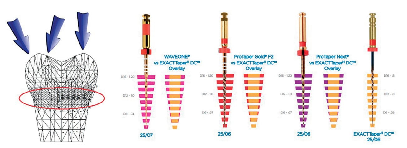
File taper determines how much dentin is removed, especially in the cervical region of the tooth. Depending on the manufacturer, files will have either a constant or a variable taper. With a constant taper, the file taper is the same as it moves from the files tip toward its shaft. A variable taper file will taper typically in the apical half, and then the remainder of the instrument either does not taper or has a lesser degree of taper. Clinically, what this means is when instrumentation is completed, a greater amount of cervical root dentin will be removed with a constant taper file than with a variable taper file, potentially weakening the cervical tooth structure.
When we compare taper between several constant taper files that are commonly used (Wave One®, ProTaper Gold, and ProTaper Next; Dentsply Sirona) and a variable taper file (ExactTaperH DC™, SS White Dental), we are able to observe that for files with the same apical size, greater amounts of dentin are removed as the cervical is approached (Figure 2). So utilization of a variable taper file aids in conservation of tooth structure and yields when instrumentation is completed. A tooth is stronger cervically due to preservation of more pericervical dentin.
When looking at the files in the ExactTaperH DC, a variable taper system, we can see that at different distances from the file’s tip, the taper varies for each of the files in the system (Figure 3). Additionally, variable taper files tend to be less stiff (more flexible) than constant taper files due to the narrower diameter of the file in the middle and coronal aspect of the fluted portion of the files. The ExactTaperH DC also by design has a 33% smaller maximum flute diameter than the constant taper files mentioned. This allows a more passive ability to follow the canal’s curvature without trying to straighten the curved canal as may present with stiffer files, providing “root form appropriate shaping.”

One potential drawback to a constant taper file is when a portion of the file engages in the canal wall or its anatomy. A “suck down” effect happens where the file threads itself further into the canal as more of the file flutes engage more canal wall, which can increase the potential for file separation.3,4 This is much less likely with a variable taper file as there is a minimized engagement of the file against the canal wall.
Endodontic phase of treatment
Following isolation of the tooth and access of the pulp chamber with identification of the canal orifices, the canals are explored with a file to establish a glide path to the working length (WL) as measured on the radiograph. When the canal is visible to the apex radiographically, the glide path (GP) rotary NiTi file (ExactTaperH DC), which is equivalent to a size 14 file with a .03 taper (Figure 1), is taken to WL to ensure a lack of obstructions is present that may hamper advancement of the subsequent files to be used. If the canal is not clear radiographically, or the GP rotary file does not advance to WL, a No. 6, 8, or 10 hand file is taken to WL and then followed up with the GP file. When the canal orifice presents as narrow or impedes advancement of the GP file or hand files, an orifice opener is useful to allow easier progression with other files. The SX orifice opener rotary NiTi file (ExactTaperH DC) is a size 15 with an .09 taper (Figure 4) and is intended to only be utilized in the coronal and middle one-third of the canal and not taken to WL.
Rotary NiTi files should be kept in constant rotational motion before entering the canal and until withdrawn from the canal to aid in prevention of the file binding in the canal. An in-and-out motion is used while brushing the canal walls while the canal system is filled with an appropriate irrigant, which aids in removal of debris within the canal while limiting potential for file binding. Should the file not be able to advance without applying apical pressure, the canal should be recapitulated with the prior used file to WL and then file size progression continued until the final file completes canal instrumentation. Most canals will be completed with an F3 ExactTaperH DC file (Figure 4), which is a size 30 with an .06 taper. Some canals — e.g., maxillary molar palatal canals, mandibular distal canals, maxillary and mandibular canines, and maxillary central incisors — may require a large file instrumentation. The F4 ExactTaperH DC file (Figure 4) with a size 40 and .06 taper is an appropriate final instrument in those canals. Should the canal be wider then the F4, the file is a loose fit. Using the F4 file with a brushing motion along the canal walls will remove any tissue and allow the irrigants to act on the canal walls in preparation for obturation. Utilizing a brushing motion is done on the out-stroke from the canal, as using that on the in-stroke may cause the file tip to bind and separate in the canal. This technique also works well in those canal shapes that are irregular or ribbon shaped in the coronal half of the canal system. Narrow canals may be found in mandibular incisors, maxillary laterals, and premolars with two canals or in older patients where some narrowing of the canals has occurred due to secondary dentin with aging. In that case, final instrumentation can be done with the F2 file (Figure 4), which has a size 25 with a .06 taper.
Instrumentation is just a part of endodontic treatment and is complemented by the obturation phase. Utilization of a single-cone obturation technique, where the gutta-percha cone matches the final file used for instrumentation, allows an intimate fit of the cone with the prepared instrumented canal minimizing the amount of sealer in the canal in comparison to gutta percha.5 With this cold technique, there is no shrinkage of the gutta percha that is found when warm obturation techniques are employed.6,7 When combined with a bioceramic sealer, which when set does not have the potential for dissolution that has been reported with ZOE- and CaOH-based sealers, the result is long-term stable endodontic treatment.8
Case 1
A 76-year-old female patient presented with pain on teeth Nos. 7 (maxillary right lateral incisor) and 11 (maxillary left canine). Clinical exam noted the coronal breakdown of both teeth without any discernable mobility. Radiographs were taken, and it was noted that tooth No. 7 presented with periapical pathology and caries connection with the pulp (Figure 5). Tooth 11 did not present with periapical pathology, but pulpal exposure was noted clinically (Figure 6). Endodontic treatment of both teeth was recommended followed by restoration with a fiber post, resin core, and full-coverage crown.

The teeth were isolated by rubber dam, and caries removed with burs and hand instruments. The canal was explored with the GP rotary file to WL. This was followed by instrumentation to the F3 ExactTaperH DC file in tooth No. 7 and the F4 ExactTaperH DC file in tooth No. 11. The canals were irrigated by alternating between NaOCl 3% (Vista Apex) and 17% EDTA solution (Vista Apex) during instrumentation and at completion. Due to the periapical pathology, it was decided to fill the canals with a CaOH medicament (Vitapex®, Neo Dental International) to the apex to allow apical healing prior to obturation of the canal systems. The teeth were sealed by placement of GC Fuji® Automix LC (GC America) as a temporary restoration until endodontic completion.
The patient returned after 2 weeks indicating all pain and sensitivity that had been present prior to treatment were completely resolved. The teeth were again isolated, and the provisional restorations removed. The canals were instrumented with the final file sizes used at the last visit and irrigated with 17% EDTA solution to remove the CaOH placed at the last appointment and dried with paper points (ExactTaperH DC) matching the size of the final file used. Bioceramic Root Canal Sealer (SS White) was mixed and dispensed on a pad. A gutta-percha cone (ExactTaperH DC) matching the final file size was coated in the sealer, and both canals were obturated in a single-cone technique. The excess cone was cut off at the canal orifice, and isolation was removed to take a final radiograph to document canal obturation (Figures 7 and 8). A temporary restoration was placed into both teeth using the GC Fuji® Automix LC, and the patient was appointed to restore the two teeth.
Case 2
A 37-year-old male new patient presented with the complaint of pain with hot foods and beverages on teeth Nos. 14 (maxillary left first molar) and 15 (maxillary second molar), which had been increasing the past 6 months since he had restorations placed by a prior dentist due to decay. The past treatment occurred before he had relocated to my area. A radiograph was taken, and slight apical widening was noted on both teeth (Figure 9). Both teeth were responsive to testing with heat and cold that lingered for a minute or two after the stimulus was removed. Both teeth also tested to slight responsiveness to percussion stimuli. The patient was informed that based on what presented, it was recommended that both teeth were moving toward needing endodontic treatment, or we could adjust the occlusion and see if that helped with symptom improvement. The patient indicated due to the increasing sensitivity to hot foods and beverages, he would prefer to initiate endodontic treatment at this time.
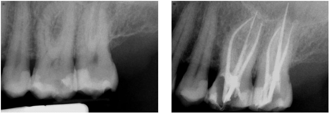
The teeth were isolated, and access performed with canal orifice identification. The SX file (ExactTaperH DC) was utilized to enlarge the canal orifice and aid in further file instrumentation. The canals were then instrumented to WL with the GP file. Each canal was instrumented with ExactTaperH DC files starting with the S1, then S2, followed by the F1, F2, and F3 in the mesial-buccal and distal-buccal canals in both molars. The palatal canals in both teeth were competed with the F4 file. The canals were irrigated by alternating between NaOCl 3% (Vista Apex) and 17% EDTA solution (Vista Apex) during instrumentation and at completion. Canals were dried with paper points (ExactTaperH DC) matching the size of the final file used. Bioceramic Root Canal Sealer (SS White) was mixed and dispensed on a pad. A gutta-percha cone (ExactTaperH DC) matching the final file size for each canal was coated with sealer, and each canal was obturated in a single-cone technique. The excess cones were cut off at the canal orifice, and obturation was completed (Figure 10). A temporary restoration was placed into both teeth using the GC Fuji® Automix LC, and the patient was appointed to restore the two teeth.
Conclusion
Endodontics has transitioned to being more conservative in preserving tooth structure and becoming restoratively driven. Long-term success can be linked to how much natural tooth structure is present following endodontic treatment in the cervical area of the tooth as under functional loading that is where stress is concentrated. A variable taper NiTi file system such as the ExactTaperH DC provides a flexible file with a minimum number of files to complete instrumentation, so a change in the practitioner’s current technique is not required. The system provides files to allow glide path formation through instrumentation with shaping and completing with finishing, plus an orifice opener when needed. The ExactTaperH DC system is paired with paper points and gutta-percha cones that correspond to the files size and taper allowing single-cone obturation to be performed. A single-cone obturation technique allows minimization of sealer in the obturation so sealer-setting shrinkage is minimized, and the single cone matching the size and shape of the final file is able to drive sealer into the adjacent canal anatomy and dentinal tubules within the canal system. When combined with the Bioceramic Root Canal Sealer, a durable seal of the canal system is achieved, and restoration of the tooth yields preservation of necessary tooth structure improving long-term overall treatment success.
Besides his success with Exact-TaperH DC and restorative endodontics, read more about Dr. Kurtzman’s practice philosophy here: https://endopracticeus.com/gregori-kurtzman-dds-magd-fpfa-facd-fadi-dicoi-dadia/
 Dr. Gregori M. Kurtzman, DDS, MAGD, FAAIP, FPFA, FACD, FADI, DICOI, DADIA, is in private general dental practice in Silver Spring, Maryland. He is a former Assistant Clinical Professor at University of Maryland in the department of Restorative Dentistry and Endodontics and a former AAID Implant Maxi-Course assistant program director at Howard University College of Dentistry. Dr. Kurtzman has lectured internationally on the topics of restorative dentistry, endodontics and implant surgery and prosthetics, removable and fixed prosthetics, and periodontics. Dr. Kurtzman has published over 760 articles globally, several ebooks, and textbook chapters. He has earned Fellowship in the AGD, American College of Dentists (ACD), International Congress of Oral Implantology (ICOI), Pierre Fauchard, ADI, Mastership in the AGD and ICOI and Diplomat status in the ICOI, American Dental Implant Association (ADIA), and International Dental Implant Association (IDIA). Dr. Kurtzman is a consultant and evaluator for multiple dental companies. He has been honored to be included in the “Top Leaders in Continuing Education” by Dentistry Today annually since 2006 and was featured on their June 2012 cover. Dr. Kurtzman can be reached at jdr_kurtzman@maryland-implants.com
Disclosure: Dr. Kurtzman has received honoraria from SS White for lectures and articles.
- Palamara D, Palamara JE, Tyas MJ, Messer HH. Strain patterns in cervical enamel of teeth subjected to occlusal loading. Dent Mater. 2000;16(6):412-419.
- Wang Q, Liu Y, Wang Z, et al. Effect of access cavities and canal enlargement on biomechanics of endodontically treated teeth: a finite element analysis. J Endod. 2020;46(10):1501-1507.
- Agarwal S, Nagpal R, Singh UP. NiTi endodontics: contemporary views reviewed. Austin J Dent. 2018; 5(4):1112.
- Diemer F, Calas P. Effect of pitch length on the behavior of rotary triple helix root canal instruments. J Endod. 2004;30(10):716-718.
- El Sayed MA, Taleb AA, Balbahaith MS. Sealing ability of three single-cone obturation systems: An in-vitro glucose leakage study. J Conserv Dent. 2013;16(6):489-493.
- Gurgel-Filho ED, Feitosa JP, Gomes BP, et al. Assessment of different gutta-percha brands during the filling of simulated lateral canals. Int Endod J. 2006;39(2):113-118.
- Lottanti S, Tauböck TT, Zehnder M. Shrinkage of backfill gutta-percha upon cooling. J Endod. 2014;40(5):721-714.
- Hegde V, Arora S. Sealing ability of three hydrophilic single cone obturation systems: An in vitro glucose leakage study. Contemp Clin Dent. 2015;6(Suppl 1):S86-S89.
Stay Relevant With Endodontic Practice US
Join our email list for CE courses and webinars, articles and more..
