Editor’s intro
Drs. Martin Trope, Klaus Lauterbach, and Gilberto Debelian pursue minimal dentin removal with adaptive core instruments.
Drs. Martin Trope, Klaus Lauterbach, and Gilberto Debelian discuss how these files clean the canal in all dimensions with minimal removal of dentin
Retreating a previously obturated root canal provides a unique set of challenges. First, the canal has already been instrumented so that the shape and remaining wall thickness is no longer in the control of the retreating practitioner. Removing the filling material can be difficult and sometimes impossible. If successful in removing the main core of filling material, as practitioners we must then disinfect the canal to remove microbes and substrate that were the cause of the failure in the first place.
Traditionally, we use the same type of files for retreatment that we have used in the initial treatment. These files have round shapes with cores that take up most of the space in the canal. The core gutta percha is softened with solvent, and then these files are used to remove the material in the same way they were used initially to do the first root canal. With most traditional files, the file core takes up the entire space provided by the canal. This creates a problem in that there is no place for the previous filling material to go. Thus, too much of the old filling material is pushed laterally in the canal, filling any available irregularity in the canal and limiting the effectiveness of any disinfectant or agitating device that comes after the core of the original filling is removed. Now, in order to remove the microbes that are the cause of the failure, the canal must be instrumented to sizes that may jeopardize the structural integrity of the root and the survivability of the tooth. Also, in some cases, where the root filling includes a carrier that cannot be softened, its removal may not be possible, necessitating a surgical approach to the case.
A previously published article in Endodontic Practice US introduces instruments with adaptive cores in order to clean the canal in all dimensions with minimal removal of dentin. (Learn more by visiting https://endopracticeus.com/clinical-articles/three-dimensional-instrumentation-reaching-next-level-endodontics.)
These instruments are the XP-3D Shaper™ and XP-3D Finisher™ (Brasseler USA®) that work together to maximally clean the canal with minimal dentin removal. The design of these instruments also makes them perfect for retreatment.
XP-3D file to start the GP removal. Once the canal is entered, the serpentine shape of the instrument will wrap around the GP and pull it out of the canal
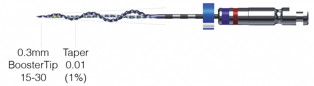
The XP-3D Shaper
The core of the Shaper file has a diameter of 0.30 mm at the tip and 0.01 taper (#30/01).
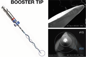
The small taper makes the file highly flexible and resistant to cyclic fatigue (Figure 1), enabling the file to be used at high speed without the fear of fracture. Speeds of 1,000 rpm to 2,500 rpm are especially useful for retreatment purposes. Critical for retreatment, the file has a Booster Tip (BT) that allows it to move into spaces as small as 15/02 or 10/04 (Figure 2).
When the initial treatment is done with round files, there is inevitably space available in the bucco-lingual dimension of the canal that is not round (Figure 3).
These non-round areas are the path that the fine tip of the Booster Tip will penetrate and proceed into the canal.
At room temperature, the file has a slight serpentine shape. When it reaches body temperature in the canal, the file stiffens enabling it to move down the canal and also cut dentin, if required. The combination of the extreme flexibility of the file plus the serpentine shape results in the file moving into the path of least resistance so that it will find the unfilled areas of the canal and move down those “least resistant” pathways.
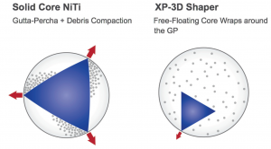
Now we have a file with a very small core diameter that will not take up the entire canal, and the serpentine shape will allow the material to be removed while staying within the “hollow” core of the file and thus not be pushed laterally in the canal.
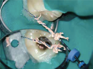
use of the XP-3D Shaper. The serpentine shape of the instrument facilitates the removal of the entire core at once. (Courtesy Dr. K. Lauterbach)
As previously mentioned, round files cannot reach non-round areas in the canal. Thus, in most of these cases, there is space in some part of the canal that has not been touched or filled (Figure 3). This space is the path of least resistance into which the Shaper will move while at the same time wrapping around the core material and pulling it out via its “empty” core (Figures 4 and 5).
From a retreatment point of view, this file is ideal for removing the previous core filling material while not creating more debris and difficulty in disinfecting the failed previous root canal (Figures 4 and 5). However it does not have the ability to clean the very irregular parts of the canal or debris and gutta percha that may be within the irregularities from the previous primary root canal instrumentation and root filling. Here the addition of the XP-3D Finisher R is required to clean the canal maximally before the canal is obturated for the second time.

in the austenite phase (bottom), the last 10 mm of the instrument achieves a sickle shape with a depth of 1.5 mm. Thus when spinning, it can reach 300, and if the sickle is compressed, the tip can expand to 600
XP-3D Finisher R (retreatment)
The XP-Finisher has a core diameter of No. 25 with no taper (00). When at body temperature, it will change shape to a sickle at the last 10 mm, giving it a capacity of No. 300 (Figure 6) when spinning and no resistance is met; but when compressed inside the canal, the tip has the capacity of up to No. 600.
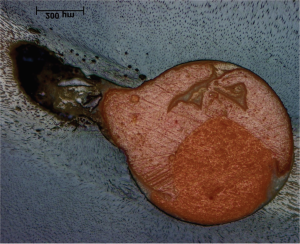
After the initial removal of the gutta-percha core material, especially with traditional files, there is inevitably gutta percha, sealer, and debris on the walls of the canal particularly around any curvature in the canal and, as already mentioned, in the irregularities of the canal (Figure 7).
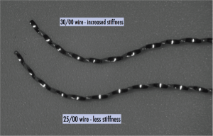
In order to more effectively remove these remnants, a XP-Finisher R has been developed. This instrument is identical to the XP-Finisher except that the core file is size 30/00 rather than size 25/00 (Figure 8). This gives the instrument more strength to dislodge sticky remnants while contacting the canal walls than the 25/00 that needs to (only) remove biofilm and microbes that should be easier to dislodge. Thus, after removal of the core gutta-percha point with the XP-3D Shaper, the XP-Finisher R is used for 60 seconds to dislodge remaining remnants that have stuck to the canal wall and irregularities. Most practitioners are amazed at the amount of debris that is seen after what they considered to be a clean canal ready for re-obturation (Figure 9).
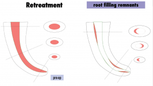
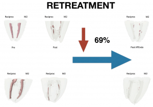
Studies have shown that the Finisher is able to remove a remarkable amount of remaining gutta percha and debris after the first attempt with traditional files. Figure 10 shows the diagrammatic results from one such study.
Case report
The following case illustrates a retreatment performed by Dr. Klaus Lauterbach using the adaptive core instruments.
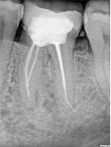
The patient presented symptomatic with a poorly filled root canal on the lower left molar with evidence of posttreatment disease.
Figure 11 shows the preoperative radiograph of the failing root canal.
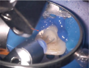
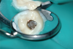
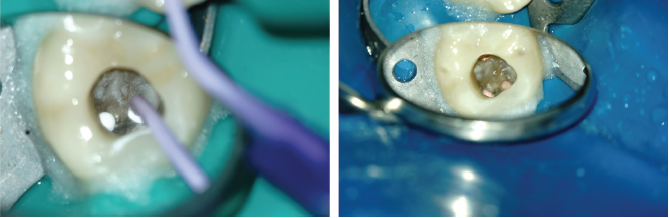
Access was prepared through the crown in order that the adaptive files could freely enter each canal (Figure 12).
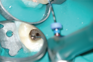
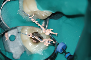
An entry path was made into the gutta-percha with a thin ultrasonic tip and the tip of the XP-3D Shaper placed into this path (Figure 13).
Using the XP-3D instrument at high speed (1,000 rpm to 2,500 rpm), the gutta percha is easily removed from the canal in thin strings wrapped around file or within the coils of the file (Figure 14).
The canal is further cleaned with the XP-Finisher R for 60 seconds, and calcium hydroxide is placed with a lentulo-spiral instrument (not shown here). Then the patient was asked to return after 1 week for obturation. The patient returned after 1 week asymptomatic, and the canal was filled with cold hydraulic obturation bioceramic GP and sealer (Figure 15).
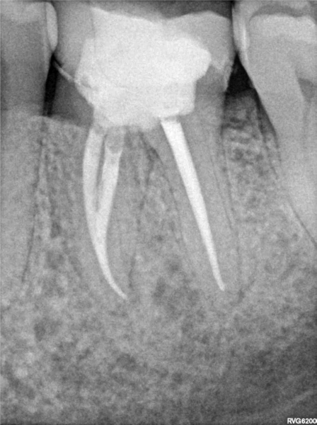
The postoperative radiograph shows a well filled root canal with evidence of sealer in the isthmus of the mesial canals (Figure 16).
Summary
Retreatment has been a challenging procedure for a number of reasons. First, the first instrumentation usually has dictated the remaining dentin in the canal that usually must be further reduced in order to adequately disinfect. Traditional instruments are poorly designed for retreatment because instead of removing old filling material, they usually spread the material into previously unfilled parts of the canal making disinfection more difficult and removal of a large amount of dentin inevitable.
The XP-3D instruments are perfectly designed for retreatment. The XP-3D Shaper is able to locate areas of least resistance due to the Booster Tip and extreme flexibility. Once a path has been found, the adaptive core of the instrument is able to extract the core gutta percha from the canal without moving it laterally, thus saving valuable dentin and maintaining the strength of the tooth before retreatment. The XP-3D Finisher R file is stiff enough to remove remaining GP, debris, and biofilm but not stiff enough to change the shape of the canal or remove noticeable dentin, thus maintaining the strength of the tooth before the root canal was started. Thus, these two instruments in combination offer tremendous advantages over the traditional round files.
Editor’s call to action
With minimal dentin removal in mind, Drs. Debelian and Trope co-authored a CE about “Cleaning the third dimension.” Read it here and subscribers can take the quiz to receive 2 CE credits!
https://endopracticeus.com/ce-articles/4948/
Stay Relevant With Endodontic Practice US
Join our email list for CE courses and webinars, articles and more..
