Editor’s intro: Dr. José Francisco Gaviño Orduña treats a permanent maxillary first molar with five canals and illustrates the complexities of the human body.
Dr. José Francisco Gaviño Orduña illustrates how 3D imaging helped during treatment of a complex tooth anatomy
Endodontists are privy to the intricate beauty that is the internal anatomy of a tooth. However, after performing hundreds of routine root canals, we may forget the surprises that can be hidden within the enamel and dentin. Such is the circumstance for one once-in-a-lifetime case that started out as “just another root canal” but ultimately reminded me of the complexities of the human body.
Patient history
A woman presented for an emergency appointment. She complained of continuous pain in the posterior upper right quadrant that was severe enough to keep her awake at night. She also experienced lingering pain, lasting more than 30 seconds, after hot or cold foods.
Exam and diagnosis
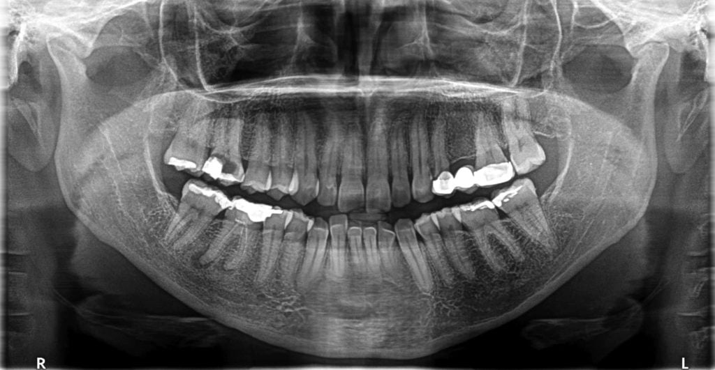
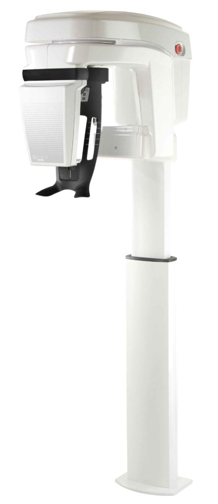
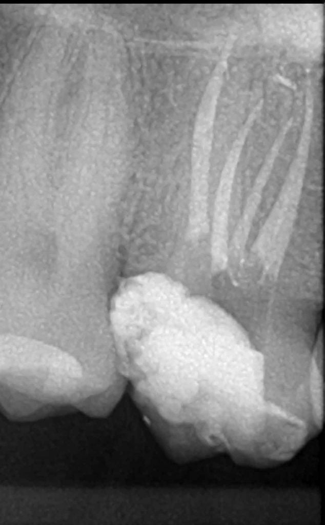
Suspecting decay, a panoramic radiograph was captured using the CS 8100 3D system (Carestream Dental) (Figure 1).
A periapical lesion was present on tooth No. 30 (FDI No. 46), and nonsurgical retreatment was offered to the patient. However, since the patient was asymptomatic, she declined treatment. Routine diagnostic tests (percussion, apical palpation, mobility, and periodontal probing) were performed and recorded for all teeth. Pulp vitality tests using thermal stimulation (cold and hot) were also performed in the upper right quadrant (area of discomfort). The patient experienced continuous pain identifying the symptomatic tooth as tooth No. 3 (FDI No. 16), describing the pain as spontaneous and lingering after the stimulus was removed. Final diagnosis was determined to be irreversible pulpitis of tooth No. 3 (FDI No. 16).
Treatment
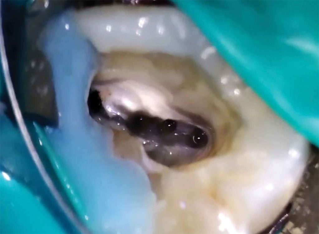
The patient then agreed to undergo root canal therapy for tooth No. 3 during the initial visit. An access cavity on tooth No. 3 was prepared with the aid of a microscope. Afterwards, using ultrasonic tips, complete root canal access was established, and five root canal orifices were discovered: three in the mesiobuccal (Figure 2), the distobuccal, and the palatal canals.
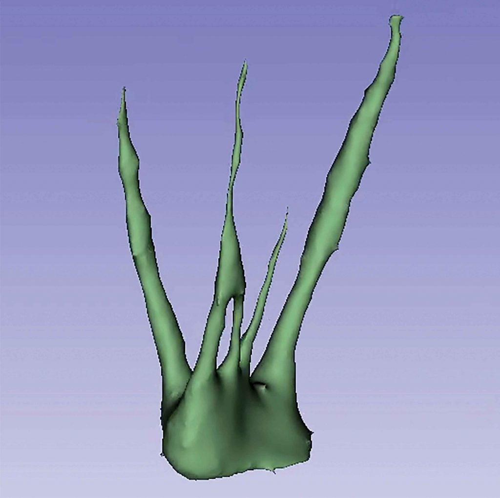
A 5 cm x 5 cm limited field of view CBCT scan was also acquired with the CS 8100 3D to gain a three-dimensional view of the tooth’s unique anatomy (Figure 3).
The oblique view of the scan confirmed the confluence of canals and allowed for better instrumentation: The mesiobuccal and mesiomiddle canals aligned perfectly with each other (Figure 4).
In dealing with this unusual situation, it was decided that the best course of action would be to treat each canal independently before they converged (Figure 5).
Following RTC, the patient returned for a third appointment for a direct overlay. The patient is now asymptomatic and may choose to return at a later date to treat the lesion found on tooth No. 30 (FDI No. 46).

Discussion
The chances of a maxillary first molar having a fifth canal is about 2%.1 However, to encounter not only a fifth canal but to see four of those canals in a perfectly straight line was a once-in-a-lifetime experience. Even as an associate professor at the University of Barcelona, who teaches students about such rare clinical situations, to encounter it in my practice was amazing.
Fortunately, the use of cone beam computed tomography allowed me to examine the unusual anatomy of the tooth in minute detail and adjust the treatment plan accordingly. The use of CBCT in endodontics in Spain is growing each year; currently, it’s more popular among oral surgeons, but there’s no doubt of its benefits to the endodontic field. The fact that treatment during the first appointment had to be stopped to take the scan highlights CBCT’s important role in treatment planning. It provides insight that microscopes and ultrasonic tips can’t the day of treatment. However, taking a CBCT scan allowed me to reevaluate treatment and move forward with confidence of greater success; since being treated in the spring of 2018, the patient has not returned complaining of pain.
This case could have gone much differently without the use of CBCT. However, much like this patient’s mesiobuccal and mesiomiddle canals, sometimes, everything lines up just perfectly for successful treatment and pain-free patients.
Besides the possibility of five canals, for more interesting tidbits about the anatomy of the root canal system, read Dr. Tony Druttman’s article, “Top ten tips: Anatomy of the root canal system” here.
 José Francisco Gaviño Orduña, DDS, earned his degree in dentistry from the University of Barcelona in 2005 and is currently studying for his PhD. He also serves as an associate professor of conservative dentistry at the University of Barcelona. Dr. Gaviño’s practice focuses on endodontics, conservative dentistry, and surgery.
- Anand P, Mahalaxmi S. Maxillary first molar with five canals. SRM J Res Dent Sci. 2016;7(1):45-47
Stay Relevant With Endodontic Practice US
Join our email list for CE courses and webinars, articles and more..
