Drs. Eugenia Pilar Consoli Lizzi, Romina Chaintiou Piorno, and Pablo Alejandro Rodríguez write that recognizing and classifying C-shaped canals can lead to more predictable outcomes.
Drs. Eugenia Pilar Consoli Lizzi, Romina Chaintiou Piorno, and Pablo Alejandro Rodríguez focus on a case showing the distinctive anatomy of C-shaped canals
Abstract
Aim
The aim of this report is to present a rare case report in which multiple C-shaped canals were diagnosed in an
adolescent male.
Case report
A healthy 16-year-old Argentine male, with no significant associated factors, was diagnosed to have six mandibular teeth with C-shaped canals by means of CBCT imaging. C-shaped canals were observed in the following teeth — mandibular right second molar, mandibular right first molar, mandibular right second premolar, mandibular left first premolar, mandibular left first molar, and mandibular left second molar — and were classified according to Fan et al. classifications. We present this report as the first published documented case of a cone-beam image diagnosed patient, exhibiting six C-shaped canals in the same jaw.
Clinical implications
Clinicians must consider the C-shaped canal anatomy in multiple presentations as a possibility in the clinical practice. Recognition and classification of C-shaped canals lead to a safer endodontic treatment and predictable outcomes.
Introduction
The main anatomical feature of the C-shaped roots is the presence of a fin or web connecting the individual canals. In molars, the canal orifice is ribbon shaped describing an arc of 180º or larger, instead of having the typical pulp chamber form with three root canals. The isthmus laying between the canals can develop difficulties when performing the canal debridement and obturation. The prevalence of C-shaped root canals is high in the mandibular second molars1; they may also appear in the mandibular first molars,2 mandibular first premolars,3,4 mandibular second pre-molars,5,6 maxillary molars,7 and even in maxillary lateral incisors.8
Ingle reported that mandibular first-premolar canal morphology is generally oval in the coronal third, round or oval in the middle third, and round in the apical third.9 On the other hand, Lu et al. stated that a circumferential or an oval canal, or two canals, could be encountered coronally, and the C-shaped cross section could be found at the apical 3 mm to 6 mm.3 The maxillary second premolar is typically described in textbooks as a single-rooted tooth with a main canal. Usually, the ovoid-shaped root cross section displays development grooves or depressions at the mesial and the distal surfaces.10 Typically, the mandibular molars exhibit two distinct roots. The mesial root feature is a flattened mesiodistal surface and a wider buccolingual surface. The distal root is mainly straight with an oval canal or two round canals.11 The C-shaped canals are more frequently observed in the mandibular second molars.12
In different research studies, Fan et al. assessed and classified the C-shaped canals (axial sections) by microcomputed tomography, and proposed two classifications.
Fan et al. classifications for mandibular second molars.13
C1: form of an uninterrupted C, with no separation or division.
C2: semicolon-shaped, C interrupted
C3c: three separate canals and C3d: two separate canals
C4: single round or oval canal
C5: absence of a root canal lumen, only seen close to the apex.
Fan et al. classification for mandibular first premolars14:
C1: form of an uninterrupted C
C2: semicolon-shaped, C interrupted
C3: two separate round-, oval-, or ribbon-shaped canals
C4: single round-, oval-, or ribbon-shaped subdivided according to Wu et al.15 (in C4a, round; C4b, oval; and C4c, ribbon-shaped)
C5: three or more separate canals
C6: absence of a root canal lumen, localized close to the apex
As a possibility, consider finding canal morphology variations before starting the treatment. Although the preoperatory radiographies provide a two-dimensional image of a three-dimensional configuration, the interpretations made by the Clark method16 could help, but would not offer detailed images in the case of complex anatomies. In contrast, the cone-beam computed tomography (CBCT) scan is a 3D-noninvasive method, which enables an accurate morphological analysis with reconstruction of dental tissues. This study reveals outer and inner anatomic details and variations, suggesting the presence of additional roots and root canals. In comparison with some other digital techniques, it offers a means for a better identification of teeth with multiple canals, such as the mandibular first premolars and the maxillary first molars.17,18
The aim of this article is to present a case report of a male patient exhibiting multiple C-shaped canals in the mandibular arch, diagnosed by CBCT imaging, and to assess and classify its morphology.
Case report
Patient
A 16-year-old Argentine male patient was referred to the School of Dentistry, University of Buenos Aires, in October 2017. A CBCT image of the full mandibular arch was taken, the scan was requested by the professionals’ team in charge of the diagnosis, and resolution of preexisting pathologies that have not been the motive of the following report. Ethical considerations were taken into account. The patient signed the informed consent form, which states that the information and the imaging studies can be utilized for academic or scientific purposes and his identity preserved by the Dentistry School (Resolution (CD) N° 983).
Medical and family/social history
The patient has no relevant medical background or record of systemic disease.
CBCT assessment
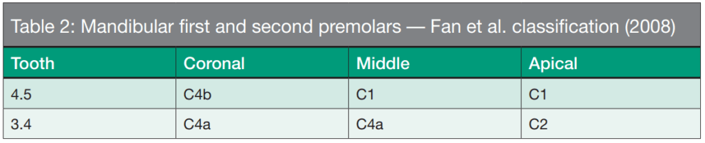
The image volume has been acquired at the Department of Diagnostic Imaging with a Kodak 9000c 3D tomograph, with 70 kV and 10 mA, exposure time of 32.4 seconds, and a voxel size of 200 µm x 200 µm x 200 µm. The DICOM data was CD recorded for its assessment. CBCT images have been assessed at the Department of Endodontics by two endodontists trained on the observation of tomography slices and updated on the knowledge of the inner dental anatomy and the identification of mandibular second molars and mandibular first premolars according to the Fan et al. classifications.13,14 In assessing the full mandibular arch volume, the impacted mandibular canines were observed. The mandibular third molars exhibited incomplete apexification. Likewise, two super-numerary teeth with incomplete apexification were impacted — one in the apical region between the mandibular right first and second premolar, and another between the mandibular right lateral incisor and the mandibular right first premolar. C-shaped canals were observed in the following teeth: mandibular right second molar, mandibular right first molar, mandibular right second premolar, mandibular left first premolar, mandibular left first molar, and mandibular left second molar (Figures 1 and 2).


The assessment was done by thirds —namely, coronal, middle and apical:
- coronal, 2 mm apical to the canal/s entrance
- apical, 2 mm above the apex
- middle, the average distance between coronal and apical
No finding has been made in the literature as it concerns the C-shaped canal classification either for mandibular second premolars or for mandibular first molars. For this reason, the four C-shaped mandibular second molars and the two C-shaped mandibular first premolars were classified according to the 2004 and 2008 Fan et al. classifications, respectively.13,14 Tables 1 and 2 show the data obtained.

 Discussion
Because of the different C-shaped canal prevalence percentages rates shown, it is thought that this case presented is unusual and interesting. Regardless of the extensive literature review, it is estimated that this is the first documented case of a cone-beam image diagnosed patient, exhibiting six C-shaped canals in the same jaw. The prevalence variations of the C-shaped canals can be explained based on the diverse assessment methodologies applied, the different sample sizes, or because of the ethnicity or the geographical region considered.
Different studies have assessed mandibular premolar root canal morphology over the years and reported that a quite high percentage of these teeth have more than one canal.4 Lu et al. assessed the mandibular first premolar canal morphology in a Chinese population and reported that 18% of the teeth configuration were C-shaped.3 Baisden et al. studied the cross sections of 106 mandibular first premolars in an American population and found that the C-shaped canal prevalence rate was 14%.4 Sikri and Sikri19 researched the morphology aberrations in the pulp space using the same method and reported that the C-shaped canal prevalence rate in a population of India was 10.7%, while Velmurugan and Sandhya20 noted a 1% prevalence rate. Yang et al.21 encountered only five cases of the 440 first premolars studied (1.14%), and Yu et al.22 found just two cases in 178 first premolars, which is a 1.1% prevalence rate. Having such a scarce number of identified samples, it is difficult to understand the relation of this anatomic condition with some other factors such as sex, localization (right or left side), and bilaterality with the aim of performing a statistical analysis. Martins et al. collected a sample of 2,012 mandibular premolars in a Portuguese population and identified 31 of them having C-shaped canals, the C-shaped configuration prevalence rate being 2.3% and 0.6% of mandibular first and second premolars, respectively.6
This prevalence decreases significantly in mandibular first molars. Some works report an incidence rate ranging from 0.85% in the Turkish23 to 0.6% in the Portuguese.24 Silva et al. observed C-shaped canals in only 12 of 460 first and second molars (2.6%); the C-shaped incidence was 4 of 234 first molars (1.7%) and 8 of 226 second molars (3.5%).25
In mandibular second molars, the highest rates in Northeast Asia were 31.5% in Chinese26 and 32.7% in Koreans,27 and a range of 2.7% to 0.6% variation is described in the Caucasian population.12,28 Considering the nationality of the patient of this case report, a study made in an Argentine subpopulation evaluating C-shaped canals in mandibular second molars by means of CBCT references an incidence of 20%.29
Conclusion
The tomographic finding in this case report is relevant because of the unusual presence of six teeth with C-shaped anatomy in the same jaw. The thorough understanding of the root anatomy complexity helps achieve a better root canal system treatment, optimizing the clinician practice.
Acknowledgments
The authors acknowledge the Department of Diagnostic Imaging for providing the CBCT image volume of the present work.
C-shaped canals are also discussed in an article by Drs. Alexandre Capelli et al. in their CE article “New resources to mitigate failure in root canal treatment and retreatment” here: https://endopracticeus.com/ce-articles/new-resources-mitigate-failure-root-canal-treatment-retreatment/
 Eugenia Pilar Consoli Lizzi, DDS, graduated from the School of Dentistry of the University of La Plata in 2013. Four years later in 2017, she received a Specialization in Endodontics from the University of Buenos Aires. Currently, Dr. Consoli Lizzi serves as an Assistant Professor in the Department of Endodontics, School of Dentistry, University of Buenos Aires, Argentine Republic.
 Romina Chaintiou Piorno, DDS, graduated from the School of Dentistry of the University of Buenos Aires in 2012. Two years later in 2014, she received a Specialization in Endodontics from the University of Buenos Aires. Currently, Dr. Chaintiou Piorno serves as an Assistant Professor in the Department of Endodontics, School of Dentistry, University of Buenos Aires, Argentine Republic.
 Pablo Alejandro Rodríguez, DDS, PhD, earned his degree in dentistry from the University of Buenos Aires in 1991. He later graduated with both a Specialization in Endodontics in 2007 and a Specialization in Prosthodontics. Dr. Rodríguez completed his PhD in 2016. Currently, he serves as the Head Professor of the Department of Endodontics and the Director of the Specialization in Endodontics. Dr. Rodríguez is also Professor of Prosthodontics in the School of Dentistry of the University of Buenos Aires. He is the actual Dean of the School of Dentistry of the University of Buenos Aires in the Argentine Republic.
Disclosure: The authors deny any conflicts of interest related to this study.
- Jafarzadeh H, Wu Y-N. The C-shaped root canal configuration: A review. J Endod. 2007;33(5):517-523.
- Bolger WL, Schindler WG. A mandibular first molar with a C-shaped root configuration. J Endod. 1988;14(10):515-519.
- Lu TY, Yang SF, Pai SF. Complicated root canal morphology of mandibular first premolar in a Chinese population using the cross section method. J Endod. 2006;32(10):932-936.
- Baisden MK, Kulild JC, Weller RN. Root canal configuration of the mandibular first premolar. J Endod. 1992;18(10):505-508.
- Cleghorn BM, Christie WH, Dong CCS. Anomalous mandibular premolars: a mandibular first premolar with three roots and a mandibular second premolar with a C-shaped canal system. Int Endod J. 2008;41(11):1005-1014.
- Martins JNR, Francisco H, Ordinola-Zapata R. Prevalence of C-shaped configurations in the mandibular first and second premolars: A cone-beam computed tomographic in vivo study. J Endod. 2017;43(6):890-895.
- Dankner E, Friedman S, Stabholz A. Bilateral C shape configuration in maxillary first molars. J Endod. 1990;16(12):601-603.
- Bóveda C, Fajardo M, Millan B. Root canal treatment of an invaginated maxillary lateral incisor with a C-shaped canal. Quintessence Int. 1999;30(10):707-711.
- Ingle JI. Endodontics. 3rd ed. Philadelphia: Lea& Febiger; 1985.
- Ingle JI, Bakland LK, Baumgartner JC. Ingle’s endodontics 6. 6th ed. Hamilton: BC Decker Inc; 2008.
- Skidmore AE, Bjorndal AM. Root canal morphology of the human mandibular first molar. Oral Surg Oral Med Oral Pathol. 1971;32(5):778-784.
- Cooke HG, Cox FL. C-shaped canal configurations in mandibular molars. J Am Dent Assoc. 1979;99(5):836-839.
- Fan B, Cheung G, Fan M, Gutmann J, Bian Z. C-shaped canal system in mandibular second molars: Part I — anatomical features. J Endod. 2004;30(12):899-903.
- Fan B, Yang J, Gutmann JL, Fan M. Root canal systems in mandibular first premolars with C-shaped root configurations. Part I: Microcomputed tomography mapping of the radicular groove and associated root canal cross sections. J Endod. 2008;34(11):1337-1341.
- Wu M-K, R’oris A, Barkis D, Wesselink PR. Prevalence and extent of long oval canals in the apical third. Oral Surg Oral Med Oral Pathol, Oral Radiol Endod. 2000;89(6):739-743.
- Clark CA. A method of ascertaining the relative position of unerupted teeth by means of film radiographs. Proc R Soc Med. 1910;3(Odontol Sect):87-90.
- Matherne RP, Angelopoulos C, Kulild JC, Tira D. Use of cone-beam computed tomography to identify root canal systems in vitro. J Endod. 2008;34(1):87-89.
- Patel S, Dawood A, Ford TP, Whaites E. The potential applications of cone beam computed tomography in the management of endodontic problems. Int Endod J. 2007;40(10):818-830.
- Sikri VK, Sikri P. Mandibular premolars: aberrations in pulp space morphology. Indian J Dent Res. 1994;5(1):9-14.
- Velmurugan N, Sandhya R. Root canal morphology of mandibular first premolars in an Indian population: a laboratory study. Int Endod J. 2009;42(1):54-58.
- Yang H, Tian C, Li G, Yang L, Han X, Wang Y. A cone-beam computed tomography study of the root canal morphology of mandibular first premolars and the location of root canal orifices and apical foramina in a Chinese subpopulation. J Endod. 2013;39(4):435-438.
- Yu X, Guo B, Li K-Z, et al. Cone-beam computed tomography study of root and canal morphology of mandibular premolars in a western Chinese population. BMC Med Imag. 2012;12(1):18.
- Demirbuga S, Sekerci Ae, Dincer An, Cayabatmaz M, Zorba Yo. Use of cone-beam computed tomography to evaluate root and canal morphology of mandibular first and second molars in Turkish individuals. Med Oral Patol Oral Cir Bucal. 2013;18(4):e737-e744.
- Martins JN, Mata A, Marques D, Caramês J. Prevalence of C-shaped mandibular molars in the Portuguese population evaluated by cone-beam computed tomography. Eur J Dent. 2016;10(3):529-535.
- Silva EJ, Nejaim Y, Silva AV, Haiter-Neto F, Cohenca N. Evaluation of root canal configuration of mandibular molars in a Brazilian population by using cone-beam computed tomography: an in vivo study. J Endod. 2013;39(7):849-852.
- von Zuben M, Martins JN, Berti L, et al. Worldwide prevalence of mandibular second molar C-shaped morphologies evaluated by cone-beam computed tomography. J Endod. 2017;43(9):1442-1447.
- Seo MS, Park DS. C-shaped root canals of mandibular second molars in a Korean population: clinical observation and in vitro analysis. Int Endod J. 2004;37(2):139-144.
- Weine FS. The C-shaped mandibular second molar: incidence and other considerations. Members of the Arizona Endodontic Association. J Endod. 1998;24(5):372-375.
- Chaintiou Piorno R, Consoli Lizzi EP, Saiegh J, et al. C-shaped canal system in mandibular second molars evaluated by cone-beam computed tomography in an Argentinean subpopulation. GJMR-J. 2019;19(4):17-23.
Stay Relevant With Endodontic Practice US
Join our email list for CE courses and webinars, articles and more..
