Drs. Prashant P. Jaju and Sushma Jaju use Orthophos SL CBCT to view commonly overlooked clinical signs of dens invaginatus.
Drs. Prashant P. Jaju and Sushma P. Jaju diagnose and treat a difficult condition with the help of smaller volume CBCT
As a practice specialized in dental radiology, many dentists use our services. We were the first practice in India to use 3D imaging. With the introduction of Dentsply Sirona’s Orthophos SL CBCT, complicated cases are diagnosed and treated efficiently and more successfully with the help of a smaller volume specific for endodontic purposes. Indeed, cone beam computed tomography is a boon for endodontists across the globe. In this article, we are presenting a difficult endodontic case in which an Orthophos SL CBCT 5 x 5.5-volume aided in identifying dens invaginatus and its subsequent treatment planning.
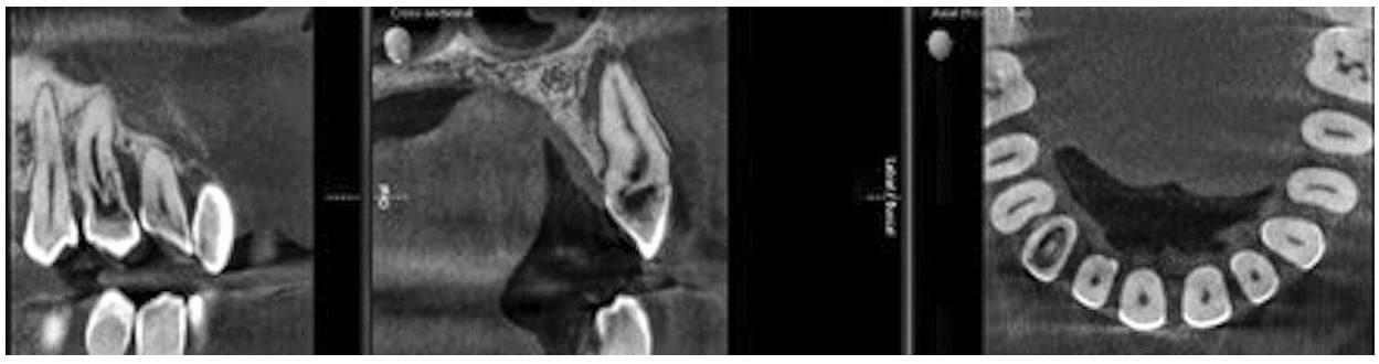
A 24-year-old male patient had swelling in the upper right canine region. An intraoral periapical radiograph showed variation in pulpal floor anatomy, but the lack of a third dimension limited its utility. For further evaluation of tooth root canal anatomy, limiting volume CBCT was advised. Orthophos SL CBCT 5 x 5.5 High Definition (HD) volume at 80 microns showed variation in pulpal floor anatomy. CBCT images revealed invagination extending through the root and communicating laterally with the periodontal ligament space through a pseudo foramen without communicating with the main root canal space. A single major orifice was present surrounded by two radiolucent areas on mesial and distal sides extending approximately 4 mm within the root not associated with the main canal (Figure 1). A single, large, periapical radiolucency was present with the tooth resulting in thinning of labial cortical plates.
This was radiographically diagnosed as a case of dens invaginatus Type IIIA, resulting in chronic periapical abscess.1 With three-dimensional visualization of the root canal space anatomy variation, the endodontist was able to proceed with a new, improved treatment protocol resulting in successful root canal filling and restoration.
Dens invaginatus is a developmental anomaly resulting in a deepening or invagination of the enamel organ into the dental papilla prior to calcification of the dental tissues. Although dens invaginatus is common, it may be easily overlooked because of the absence of any significant clinical signs of the anomaly.
Periapical radiographs are limited in revealing the type, extension, and complex morphology of dens invaginatus as well as the actual bone loss when compared to tomographic techniques. More advanced imaging techniques such as CBCT may aid the diagnosis as well as the management plan and follow-up of teeth with this developmental defect.2
Read more about the versatility of Orthophos SL CBCT in our sister publication, Orthodontic Practice US: https://orthopracticeus.com/industry-news/dentsply-sirona-imaging-enhances-versatility-orthophos-sl/
- Alani A, Bishop K. Dens invaginatus. Part 1: Classification, prevalence and aetiology. Int Endod J. 2008;41(12): 1123-1136.
- Pradeep K, Charlie M, Kuttappa MA, Rao PK. Conservative management of Type III dens in dente using cone beam computed tomography. J Clin Imaging Sci. 2012;2(1): 51.
This article was previously published in the brochure “Orthophos SL – around the world,” 2019 by Dentsply Sirona.
Stay Relevant With Endodontic Practice US
Join our email list for CE courses and webinars, articles and more..
