Editor’s intro: BeamReaders team of more than 50 oral and maxillofacial radiologists can be a formidable defense against deficient interpretations of CBCT scans.
Drs. Poorya Jalali and Mehrnaz Tahmasbi discuss how having the trained eye of a radiologist can offer peace of mind to diagnostics
 As the role of CBCT imaging continues to grow in the specialty of endodontics, a BeamReaders® Oral and Maxillofacial Radiologist is an essential partner for your practice. Integrating a radiologist into your CBCT workflow is the best defense against deficient interpretations — helping ensure optimum outcomes for your patients and a reduction in your liability related to 3D imaging.
Satisfaction of search bias
Clinicians utilize CBCT scans to answer specific clinical questions, such as, “Is there a periapical radiolucency present?” Some may be so focused on answering the original question that additional findings, which may be present, may be overlooked. Satisfaction of search bias occurs when an abnormal radiographic finding (e.g. periapical radiolucency) is detected, leading clinicians to believe their search is over. As such, the additional findings (e.g. invasive cervical root resorption of the adjacent tooth) may be missed. This is where a BeamReaders radiologist plays a valuable role.
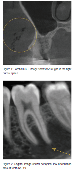
A radiologist is trained to thoroughly and interactively examine the multiplanar images in three dimensions and provide a comprehensive evaluation of the study. This structured method of evaluating a CBCT scan minimizes the risk of missing an important incidental finding. For example, Figure 1 shows foci of gas in the right buccal space, consistent with subcutaneous emphysema. This incidental finding was detected by a radiologist when evaluating a CBCT obtained for endodontic evaluation.
Availability bias
Another bias that can contribute to an error in interpreting CBCT studies is availability bias. Availability bias occurs when clinicians rely on easily recalled examples when making a diagnosis, therefore preventing them from considering alternative options. As an old saying goes, “To a man with a hammer, everything looks like a nail.” This type of bias is well-known in radiology and can best be avoided when radiographic and clinical findings are correlated, and the most probable etiologies are considered in the differential diagnosis. Figure 2 shows a periapical radiolucency similar to an endodontic pathosis. However, the endodontist noticed a normal response to sensibility testing. Correlated with the clinical testing, the radiologist considered the traumatic bone cyst foremost in the differential diagnosis, which was then confirmed by exploratory surgery.
With busy practices, practitioners don’t have the significant time to fully examine and document each CBCT study. The reports crafted by BeamReaders radiologists illustrate the clinical findings, reduce the dentists’ liability, and save clinical time. The provided information in the CBCT report includes, but is not limited to, periapical pathosis, dental anomalies, accessory/ anomalous canals, root morphology, root fracture, root resorption, and surgical planning. The latter includes information regarding the size and location of a lesion or root apex in relation to vital structures and the presence of an accessory neurovascular canal in proximity to the region of interest before a surgical procedure. A BeamReaders report provides a great platform to easily educate the patient, engage other treatment providers, increase confidence in treatment planning, and reduce associated liability. These benefits will invariably help improve outcomes and efficiency, thus helping to strengthen the reputation and productivity of an endodontic practice.
About BeamReaders®
BeamReaders is a team of more than 50 Oral and Maxillofacial Radiologists working to help you succeed. Submit your first case for free today by registering at www.BeamReaders.com and using the registration code BR4ENDO.
Editor’s call to action
Read more about how BeamReaders can provide a defense against deficient interpretations of CBCT imaging in Dr. Andrew Trosien’s article, “An introduction to reviewing CBCT images.”
https://www.orthopracticeus.com/free-ce/an-introduction-to-reviewing-cbct-images.
Stay Relevant With Endodontic Practice US
Join our email list for CE courses and webinars, articles and more..

