Dr. John Rhodes looks at locating canal orifices
Modern preparation techniques provide a very efficient means of rapidly tapering the primary root canals prior to disinfection, but in practice, the first hurdle is often being able to actually find the orifices. An infected, missed canal could result in a persistent inflammatory response and failure of treatment.
This article describes how to decode the pulp floor map in order to locate difficult to find canal orifices.
An infected, missed canal
could result in a
persistent inflammatory
response and failure
of treatment.
Decoding the pulp floor map
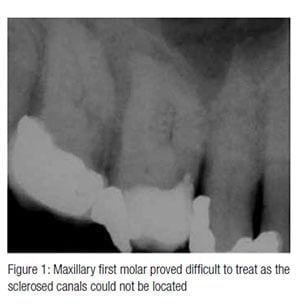
 The maxillary first molar in Figure 1 proved difficult to root treat as the sclerosed canals could not be located. The following steps demonstrate how to make the process more achievable.
Good access
The access cavity must be located correctly and provide sufficient space to allow adequate visualization of the pulp floor. This means removing tooth substance conservatively, but not compromising the ability to work efficiently during mechanical preparation.
By removing the entire restoration in this case, orientation and visualization of the pulp floor was maximized. The same is true if the operator has created an oversized access in a previous attempt at root treatment; this can be used to advantage without affecting the integrity of the tooth.
Magnification and illumination
Magnification and illumination are essential. A microscope provides the best operating field, and this is why endodontists routinely use them. Being able to see where you are working is a massive advantage, and many of the instruments require direct vision in order to be used safely.
The pulp floor
The pulp floor tends to be darker than the walls of the access cavity, but when irritation dentin is present in the pulp chamber, this can be difficult to interpret. Calcified material needs to be removed with either ultrasonic instruments or a bur such as the Tungsten carbide LN bur (Dentsply).
To prevent perforation of the pulp floor, an estimation of safe depth can be made from the preoperative radiograph, and direct vision with magnification and illumination will allow instruments to be used safely while refining the access and pulp floor. The canal orifices tend to be located at the extremities of the darker pulp floor and may appear as a small white dot if packed with dentin chips.
Use a micro-opener (Dentsply) to gauge for signs of an orifice. In a maxillary molar such as the one in this article, the palatal canal is likely to be most readily located and, once confirmed, working along the border of the pulp floor map makes location of the main buccal canals easier.
In this case, an attempt had already been made to locate the canals; the divots and iatrogenic irregularities created by burs can be disorientating, so great care is required to establish and confirm the true pulp floor map to allow identification of the canal orifices.
In the maxillary first molar, there is often a lip of dentin covering the second mesiobuccal canal that needs to be removed in order to locate the orifice. Once the primary mesiobuccal canal has been located, look for signs of an isthmus — following this in the direction of the palatal canal will often lead you to the second canal.
Ultrasonics
There are many ultrasonic instruments available for working deep in the access cavity to remove calcifications and trough between canals; some are diamond coated while others have machined tips.
In this case, a No. 3 Start-X™ (Dentsply) instrument was used, vibrated at medium power in a Piezo ultrasonic unit (Satelec, Acteon). The tip can be used dry or with water spray, but it is important to be able to see where the tip is cutting to avoid perforation. Dentin chips and smear can be washed away with sodium hypochlorite, EDTA, or citric acid to clear the operative field.
Gaining access
The tip of a fine file, DG16 endodontic probe, or micro-opener (Dentsply) will catch in the orifice of the canal once located. This can be teased open with the micro-opener before confirming patency in the coronal aspect with a flexible hand file and continuing with preparation.
Watch the video
To see how these steps are applied, visit https://youtu.be/BL2MKxF4vK0 or search
Youtube for “Endo Practice — Decoding the pulp floor map.” The author is happy to
answer questions directly via Youtube or Twitter @johnrhodesendo.
Stay Relevant With Endodontic Practice US
Join our email list for CE courses and webinars, articles and more..

 John Rhodes, BDS(Lond), FDS RCS(Ed), MSc, MFGDP(UK), MRD RCS(Ed), is a specialist in endodontics, the author of textbooks and numerous papers, and owner of The Endodontic Practice, Poole and Dorchester. He lectures and teaches on endodontics throughout the United Kingdom.