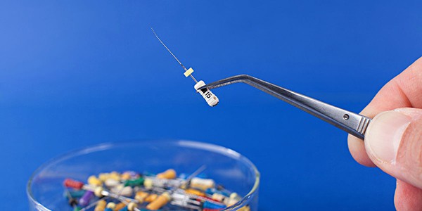
Dr. Kishor Gulabivala presents the latest literature, keeping you up-to-date with the most relevant research
Predicting severe pain after root canal therapy in the National Dental PBRN
Law AS, Nixdorf DR, Aguirre AM, Reams GJ, Tortomasi AJ, Manne BD, Harris DR, National Dental PBRN Collaborative Group. Journal of Dental Research. (0015) 94(3 Suppl):37S-43S
Abstract
Aim: Some patients experience severe pain following root canal therapy (RCT) despite advancements in care. The aim of this study was to identify factors that can be measured preoperatively, which predict this negative outcome so that future research may focus on pre-emptive steps to reduce postoperative pain intensity.
Methodology: Sixty-two practitioners (46 general dentists and 16 endodontists) who are members of the National Dental Practice-Based Research Network (PBRN) enrolled patients receiving RCT for this prospective observational study. Baseline data collected from patients and dentists were obtained before treatment. Severe postoperative pain was defined based on a rating of greater than seven on a scale from zero (no pain) to 10 (pain as bad as can be) for the worst pain intensity experienced during the preceding week, and this was collected 1 week after treatment. Multiple logistic regression analyses were used to develop and validate the model.
Results: A total of 708 patients were enrolled during a 6-month period. Pain intensity data were collected 1 week post-operatively from 652 patients (92.1%), with 19.5% (n = 127) reporting severe pain. In multivariable modeling, baseline factors predicting severe postoperative pain included current pain intensity (odds ratio [OR], 1.15; 95% confidence interval [CI], 1.07 to 1.25; P = 0.0003), number of days in the past week that the subject was kept from their usual activities due to pain (OR, 1.32; 95% CI, 1.13 to 1.55; P = 0.0005), pain made worse by stress (OR, 2.55; 95% CI, 1.22 to 5.35; P = 0.0130), and a diagnosis of symptomatic apical periodontitis (OR, 1.63; 95% CI, 1.01 to 2.64; P = 0.0452). Among the factors that did not contribute to predicting severe postoperative pain were the dentist’s specialty training, the patient’s age and sex, the type of tooth, the presence of swelling, or other pulpal and apical endodontic diagnoses.
Conclusions: Factors measured pre-operatively were found to predict severe postoperative pain following RCT. Practitioners could use this information to better inform patients about RCT outcomes and possibly use different treatment strategies to manage their patients.
Effect of education intervention on the quality and long-term outcomes of root canal treatment in general practice
Koch M, Wolf E, Tegelberg A, Petersson K. International Endodontic Journal. (2015) 48(7):680-9
Abstract
Aim: To compare the technical quality and long-term outcomes of root canal treatment by general practitioners of a Swedish Public Dental Service, before and after an endodontic education, including Ni-Ti rotary technique (NiTiR).
Methodology: A random sample was compiled, comprising one root-filled tooth from each of 830 patients, treated by 69 general practitioners participating in the education: 414 teeth root-filled in 2002, pre-education, using primarily stainless steel instrumentation and filling by lateral compaction, and 416 teeth root filled post-education (2005), using mainly NiTiR and single-cone obturation. Follow-up radiographs taken in 2009 were evaluated alongside immediate post-filling radiographs from 2002 to 2005. The density and length of the root fillings were registered; periapical status was assessed by the Periapical Index (PAI), using two definitions of disease: apical periodontitis (AP) (PAI 3 + 4 + 5) and definite AP (PAI 4 + 5). Tooth survival was registered. Root fillings pre- and post-education were compared using chi-square and Fisher’s exact tests. Crude extraction rates per 100 years were calculated for comparison of tooth survival. Explanatory variables (type of tooth, root-filling quality, periapical status, marginal bone loss, type and quality of coronal restoration) in relation to the dependent variable (AP at follow-up) were analyzed by multivariable logistic regression.
Results: Follow-up data were available for 229 (55%) of teeth treated pre-education and 288 (69%) treated post-education: Both tooth survival (P < 0.001) and root-filling quality were significantly higher (P < 0.001) in the latter. However, there was no corresponding improvement in periapical status. Both pre- and post-education root fillings with definite AP on completion of treatment had significantly higher odds of AP or definite AP at follow-up. For teeth treated post-education, inadequate root-filling quality was significantly associated with AP at follow-up.
Conclusions: Despite a higher tooth survival rate and a significant improvement in technical quality of root fillings after the education, there was no corresponding improvement in periapical status.
Mesenchymal dental pulp cells attenuate dentin resorption in homeostasis
Zheng Y, Chen M, He L, Marao HF, Sun DM, Zhou J, Kim SG, Song S, Wang SL, Mao JJ. Journal of Dental Research. (2015) 94(6):821-7
Abstract
Aim: Dentin in permanent teeth rarely undergoes resorption in development, homeostasis, or aging, in contrast to bone that undergoes periodic resorption/remodeling. The authors hypothesized that cells in the mesenchymal compartment of dental pulp attenuate osteoclastogenesis.
Methodology: Mononucleated and adherent cells from donor-matched rat dental pulp (dental pulp cells [DPCs]) and alveolar bone (alveolar bone cells [ABCs]) were isolated and separately co-cultured with primary rat splenocytes. In vivo, rat maxillary incisors were atraumatically extracted (without any tooth fractures), followed by retrograde pulpectomy to remove DPCs and immediate replantation into the extraction sockets to allow repopulation of the surgically treated root canal with periodontal and alveolar bone-derived cells.
Results: Primary splenocytes readily aggregated and formed osteoclast-like cells in chemically defined osteoclastogenesis medium with 20 ng/mL of macrophage colony-stimulating factor (M-CSF) and 50 ng/mL of receptor activator of nuclear factor kappa-b ligand (RANKL). Strikingly, DPCs attenuated osteoclastogenesis when co-cultured with primary splenocytes, whereas ABCs slightly but significantly promoted osteoclastogenesis. DPCs yielded ~20-fold lower RANKL expression but >2-fold higher osteoprotegerin (OPG) expression than donor-matched ABCs, yielding a RANKL/OPG ratio of 41:1 (ABCs:DPCs). Vitamin D3 significantly promoted RANKL expression in ABCs and OPG in DPCs. In the vivo experiment, after 8 weeks, multiple dentin/root resorption lacunae were present in root dentin with robust RANKL and OPG expression. There were areas of dentin resorption alternating with areas of osteodentin formation in root dentin surface in the observed 8 weeks.
Conclusions: These findings suggest that DPCs of the mesenchymal compartment have an innate ability to attenuate osteoclastogenesis, and that this innate ability may be responsible for the absence of dentin resorption in homeostasis. Mesenchymal attenuation of dentin resorption may have implications in internal resorption in the root canal, pulp/dentin regeneration, and root resorption in orthodontic tooth movement.
A prospective study of the incidence of asymptomatic pulp necrosis following crown preparation
Kontakiotis EG, Filippatos CG, Stefopoulos S, Tzanetakis GN. International Endodontic Journal. (2015) 48(6):512-7
Abstract
Aim: To determine the incidence of asymptomatic pulp necrosis following crown preparation as well as the positive predictive value of the electric pulp testing.
Methodology: A total of 120 teeth with healthy pulps scheduled to receive fixed crowns (experimental teeth) were included. Teeth were divided into two groups according to the preoperative crown condition (intact teeth and teeth with preoperative caries, restorations or crowns) and into four groups according to tooth type (maxillary anterior teeth, maxillary posterior teeth, mandibular anterior teeth, and mandibular posterior teeth). Experimental and control teeth were submitted to electric pulp testing on three different occasions before treatment commencement (stage zero), at the impression-making session (stage one) and just before the final cementation of the crown (stage two). Teeth that were considered to contain necrotic pulps were submitted to root canal treatment. Upon access, absence of bleeding was considered as a confirmation of pulp necrosis. Data were analyzed using bivariate (chi-square) and multivariate analysis (logistic regression). All reported probability values (P-values) were based on two-sided tests and compared to a significance level of 5%.
Results: The overall incidence of pulp necrosis was 9%. Intact teeth had a significantly lower incidence of pulp necrosis (5%) compared with preoperatively structurally compromised teeth (13%) [(OR: 9.113, P = 0.035)]. No significant differences were found among the four groups with regard to tooth type (P = 0.923). The positive predictive value of the electric pulp testing was 1.00.
Conclusions: The incidence of asymptomatic pulp necrosis of teeth following crown preparation is noteworthy. The presence of preoperative caries, restorations or crowns of experimental teeth correlated with a significantly higher incidence of pulp necrosis. Electric pulp testing remains a useful diagnostic instrument for determining the pulp condition.
Three-year outcomes of root canal treatment: Mining an insurance database
Raedel M, Hartmann A, Bohm S, Walter MH. Journal of Dentistry. (2015) 43(4):412-7
Abstract
Aim: There is doubt whether success rates of root canal treatments reported from clinical trials are achievable outside of standardized study populations. The aim of this study was to analyze the outcome of a large number of root canal treatments conducted in general practice.
Methodology: The data were collected from the digital database of a major German national health insurance company. All teeth with complete treatment data were included. Only patients who had been insurance members for the whole 3-year period from 2010 to 2012 were eligible. Kaplan-Meier survival analyses were conducted based on completed root canal treatments. Target events were re-interventions such as (1) retreatment of the root canal treatment, (2) apical root resection (apicoectomy), and (3) extraction. The influences of vitality status and root numbers on survival were tested with the log-rank test.
Results: A total of 556,067 root canal treatments were included. The cumulative overall survival rate for all target events combined was 84.3% for 3 years. The survival rate for nonvital teeth (82.6%) was significantly lower than for vital teeth (85.6%; p < 0.001). The survival rate for single-rooted teeth (83.4%) was significantly lower than for multi-rooted teeth (85.5%; p < 0.001). The most frequent target event was extraction followed by apical root resection and retreatment.
Conclusions: Based on these 3-year outcomes, root canal treatment is considered a reliable treatment in practice routine under the conditions of the German national health insurance system. Root canal treatment can be considered as a reliable treatment option suitable to salvage most of the affected teeth. This statement applies to treatments that in the vast majority of cases were delivered by general practitioners under the terms and conditions of a nationwide health insurance system.
Outcome of nonsurgical retreatment and endodontic microsurgery: a meta-analysis
Kang M, In Jung H, Song M, Kim SY, Kim HC, Kim E. Clinical Oral Investigations. (2015) 19(3):569-82
Abstract
Aim: The purpose of this study was to evaluate and compare the clinical and radiographic outcomes of nonsurgical endodontic retreatment and endodontic microsurgery by a meta-analysis.
Methodology: Electronic databases, including Pubmed, Embase, Medline, and the Cochrane Library, were searched, and the references of related articles were manually searched to identify all the clinical studies that evaluated the clinical and radiographic outcomes after retreatment or microsurgery. The first- and second-screening processes were conducted by three reviewers independently. The final studies were selected after strict application of the inclusion and exclusion criteria. The random effects meta-analysis model with the DerSimonian-Laird pooling method was performed. The weighted pooled success rates, and 95% confidence interval estimates of the outcome were calculated. Additionally, the effects of the follow-up period and study quality were investigated by a subgroup analysis.
Results: Endodontic microsurgery and nonsurgical retreatment have stable outcomes presenting 92% and 80% of overall pooled success rates, respectively. The microsurgery group had a significantly higher success rate than the retreatment group. When the data were organized and analyzed according to their follow-up periods, a significantly higher success rate was found for the microsurgery group in the short-term follow-up (less than 4 years), whereas no significant difference was observed in the long-term follow-up (more than 4 years).
Conclusions: Endodontic microsurgery was confirmed as a reliable treatment option with favorable initial healing and a predictable outcome. Clinicians may consider the microsurgery as an effective way of retreatment as well as nonsurgical retreatment depending on the clinical situations.
Analysis of the possible causes of endodontic treatment failure by inspection during apical micro-surgery treatment
Qian WH, Hong J, Xu PC. Shanghai Kou Qiang Yi Xue/Shanghai Journal of Stomatology. (2015) 24(2):206-9
Abstract
Aim: To analyze the possible causes of previous endodontic treatment failure by microscopic inspection during apical microsurgery.
Methodology: Two hundred and eighty-nine teeth of previous endodontic treatment failure were collected from patients in Shanghai Xuhui District Dental Center between January 2006 and January 2014. All surgical procedures were performed by using an operating microscope, and 238 roots were included in the study. The surface of the apical root to be resected or the resected root surface after methylene blue staining was examined during the surgical procedure and inspected with 26 magnification to determine the state of the previous endodontic treatment by using an operating microscope. Fisher’s exact test was used to analyze the data with SPSS 19.0 software package.
Results: Among the 238 roots with periapical surgery, analysis of the reasons for previous endodontic treatment failure included leaky canal (29.41%), missing canal (15.55%), underfilling (15.55%), anatomical complexity (7.98%), overfilling (4.20%), apical fenestration (4.20%), iatrogenic problem (3.36%), apical calculus (2.52%), apical cracks (1.68%), and unknown reasons (15.55%). The frequency of possible failure causes and tooth position was closely correlated (p < 0.001).
Conclusions: Apical microsurgery can better inspect possible causes of previous endodontic treatment failure in order to improve the success rate of endodontic treatment.
Evaluating the effects of different dental devices on implantable cardioverter defibrillators
Maheshwari KR; Nikdel K; Guillaume G; Letra AM; Silva RM; Dorn SO. Journal of Endodontics. (2015) 41(5):692-5
Abstract
Aim: The implantable cardioverter defibrillator (ICD) is an electronic device that emits electrical signals to the heart via lead wires and electrodes. It is used for cardiac rhythm monitoring and treatment. Because electronic dental devices have been shown to produce electromagnetic fields, we hypothesize that they may interfere with ICD function.
Methodology: Nine dental devices (heat carrier, electronic apex locator, electric pulp tester, unipolar electrosurgery unit, electric motor, curing light, and three gutta-percha guns) were tested in this study for their ability to interfere with the function of four ICDs (two single-chambered and two dual-chambered ICDs). ICD activity was monitored for 30 seconds using an ICD programmer (Medtronic 2090; Minneapolis, Minnesota) and evaluated through an electrogram test strip printout.
Results: Electromagnetic interference was detected with the electric motor, curing light, electric pulp tester, and electrosurgery unit, although no electromagnetic disturbances were detected with these devices. No electromagnetic interferences were observed for the gutta-percha guns, heat carrier, and apex locator. However, the electro-surgery unit affected the dual-chambered ICD (Consulta CRT-D, Medtronic) and delivered therapies for fibrillation when no ventricular fibrillation was present.
Conclusions: Our results suggest that the electrosurgery unit produces electro-magnetic disturbances with unwanted therapy delivery shock and potentially clinically significant outcomes.
Dental pulp: correspondences and contradictions between clinical and histological diagnosis
Giuroiu CL, Caruntu ID, Lozneanu L, Melian A, Vataman M, Andrian S. Biomed Research International. (2015) 2015:960321
Abstract
Aim: Dental pulp represents a specialized connective tissue enclosed by dentin and enamel — the most highly mineralized tissues of the body. Consequently, the direct examination as well as pathological evaluation of dental pulp is difficult. Within this anatomical context, our study aimed to evaluate the correlation between dental pulp lesions and clinical diagnosis.
Methodology: Pulpectomies were performed for 54 patients with acute and chronic irreversible pulpitides and for five patients (control group) with orthodontic extractions. The morphological features were semi-quantitatively assessed by specific score values. The clinical and morphological correspondence was noted for 35 cases (68.62%), whereas inconsistency was recorded for 16 cases (31.38%).
Results: The results of the statistical analysis revealed the correlations between clinically and pathologically diagnosed acute/chronic pulpitides. No significant differences were established between the score values for inflammatory infiltrate intensity, collagen depositions, calcifications and necrosis, and acute, respectively, chronic pulpitides. We also obtained significant differences between acute pulpitides and inflammatory infiltrate and calcifications and between chronic pulpitides and inflammatory infiltrate, collagen deposition, and calcifications.
Conclusions: On the basis of the predominant pathological aspects — namely, acute and chronic pulpitis — we consider that the classification schemes can be simplified by adequately reducing the number of clinical entities.
Pathways of fear and anxiety in endodontic patients
Carter AE, Carter G, George R. International Endodontic Journal. (2015) 48(6): 528-32
Abstract
Aim: To evaluate the most common pathways of fear and anxiety in patients who have had root canal treatment or are planned to have one.
Methodology: Six hundred and twenty-seven patients were approached to participate of which 594 patients (20-90 years) accepted. All consenting patients had a root filling or were treatment planned to have one. The survey by Ost and Hugdahl on anxiety response patterns was modified and used. Data were presented using descriptive statistics, tested for normality using the Kolmogorov-Smirnov test and analyzed with nonparametric anova (Kruskal-Wallis) and post hoc test.
Results: Cognitive conditioning and parental pathways seem to be the primary cause (p < 0.05) of fear and anxiety with root canal treatment. Females were significantly more likely to be influenced by indirect conditioned experiences such as informative, parental, verbal threat, and vicarious pathways.
Conclusion: The origin of patients’ fears requires more attention in terms of treating endodontic-related fear and anxiety. More detailed research into the effects of demographics, causative factors, and ethnicity on pathways of fear in dentistry is required to help dentists better manage patients in a multicultural society.
Stay Relevant With Endodontic Practice US
Join our email list for CE courses and webinars, articles and more..


